Targeting BRAF V600E: Using A375 Series Cells to Evaluate Inhibitor Efficacy
Introduction:
Malignant melanoma represents the skin cancer type with the highest mortality rate. Approximately 50% of patients carry BRAF gene mutations, with the V600E mutation being the most prevalent. This mutation leads to constitutive activation of the MAPK signaling pathway, driving the malignant proliferation of tumor cells. The A375 cell line, derived from a 54-year-old female with malignant melanoma, serves as a core model for developing targeted drugs like Vemurafenib and Dabrafenib due to its endogenous BRAF V600E mutation. However, the prevalent issue of acquired resistance in clinical settings demands higher standards for preclinical research. To precisely monitor the dynamic processes of tumor regression and relapse, researchers developed A375-luc cell (bioluminescence-labeled) and A375-LUC-GFP (dual-labeled) variants. This article details how to utilize these cell lines to construct a complete evaluation system, ranging from in vitro screening to in vivo mechanism analysis.
Endogenously harboring the BRAF V600E mutation, the A375 cell line is the premier in vitro model for screening Vemurafenib and MEK inhibitors. Shop now>>
Target Validation: Sensitivity to MAPK Pathway Inhibitors
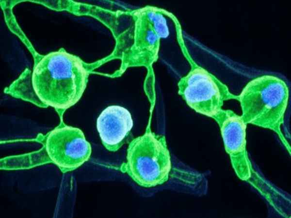
Establishing the baseline in vitro sensitivity of A375 cells to specific drugs is a necessary step before constructing complex animal models. The A375 cell line exhibits typical epithelioid morphology and maintains rapid proliferation in vitro.
Due to the presence of the BRAF V600E mutation, A375 cells rely heavily on the MAPK/ERK signaling pathway for survival. In drug screening experiments, treating parental A375 cells with varying concentrations of BRAF inhibitors (e.g., PLX4032) typically results in significant cell cycle arrest and apoptosis induction. Dose-response curves generated via CCK-8 or MTS assays allow for the determination of the half-maximal inhibitory concentration (IC50). This data validates the biological activity of the drug and provides a reference for dosage design in subsequent in vivo experiments. For combination therapy studies, such as combining with MEK inhibitors (Trametinib), the A375 model also sensitively reflects synergistic lethal effects between drugs.
In Vivo Efficacy: Quantitative Advantages of Bioluminescence Imaging
Traditional xenograft models often employ subcutaneous inoculation, relying on caliper measurements of tumor length and width to calculate volume. However, melanoma tissues treated with drugs often undergo liquefactive necrosis or fibrosis, causing volume measurements to fail in accurately reflecting the number of viable tumor cells.
Utilizing the A375-luc cell to construct tumor-bearing mouse models effectively resolves this issue. This cell line stably expresses Firefly Luciferase. Upon injection of the substrate D-Luciferin, living cells generate light signals via an ATP-dependent enzymatic reaction. The Total Flux (p/s) detected by IVIS systems correlates strictly linearly with the number of metabolically active tumor cells. In efficacy evaluations, the rapid decline of light signals in the treatment group provides intuitive evidence of drug-induced tumor regression, offering significantly higher sensitivity than volume measurement and enabling the detection of residual minimal lesions.
Utilize the A375-luc cell for long-term longitudinal monitoring to precisely capture the critical time window when resistant melanoma shifts from remission to relapse. Get a look>>
Resistance Tracking: Capturing the Relapse Window
Acquired resistance is a primary obstacle in the clinical application of BRAF inhibitors. Simulating this process in animal models requires long-term longitudinal monitoring of tumors.
Using the A375-luc cell, researchers can continuously observe single mice for weeks or even months. Typical resistance induction experiments involve continuous administration of low-to-moderate doses of inhibitors. Initially, the bioluminescent signal decreases significantly, entering a "plateau" or "remission phase." However, as resistant clones are selected and enriched, the light signal rises exponentially again. The turning point of this "U-shaped" growth curve precisely marks the time window of tumor relapse. Capturing this critical node is vital for studying the kinetic characteristics of resistance development.
Mechanism Analysis: Precise Sorting via Dual-Modal Labeling
Upon tumor relapse, parsing the molecular mechanism of resistance typically requires extracting tumor cells for sequencing. However, solid tumor tissues contain a mixture of abundant host-derived stromal cells (such as murine fibroblasts, vascular endothelial cells, and immune cells), which severely interfere with gene expression analysis of human tumor cells.
Introducing the A375-LUC-GFP dual-labeled cell line is an effective strategy to address this problem. At the experimental endpoint, resected resistant tumor tissues are digested into single-cell suspensions. Using Green Fluorescent Protein (GFP) as a selection marker, researchers can precisely separate human tumor cells (GFP-positive) from murine stromal cells (GFP-negative) via Fluorescence-Activated Cell Sorting (FACS). Subsequent transcriptomic (RNA-seq) or proteomic analysis of the purified resistant A375 strain can clearly identify bypass activation mechanisms (such as NRAS mutations or CRAF overexpression), thereby guiding the development of second-line treatment regimens.
Leverage the green fluorescent marker of A375-LUC-GFP to easily eliminate host stromal interference, isolating high-purity tumor cells for resistance mechanism analysis. Learn more>>
References
[1]Giard, D. J., et al. (1973). In vitro cultivation of human tumors: establishment of cell lines derived from a series of solid tumors. Journal of the National Cancer Institute, 51(5), 1417-1423.
[2]Davies, H., et al. (2002). Mutations of the BRAF gene in human cancer. Nature, 417(6892), 949-954.
[3]Jenkins, D. E., et al. (2005). Bioluminescent human breast cancer cell lines that permit rapid and sensitive in vivo detection of mammary tumors and multiple metastases in immune-deficient mice. Breast Cancer Research, 7(4), R444.
[4]Smalley, K. S., et al. (2006). Multiple signaling pathways must be targeted to overcome drug resistance in cell lines derived from melanoma metastases. Oncogene, 25(34), 4706-4716.
