A549-Luc: Engineering a High Bone-Metastatic Lung Adenocarcinoma Model for Metastasis Mechanism Studies
Introduction
Bone is one of the most common metastatic sites for lung adenocarcinoma, occurring in 30–40% of advanced non-small cell lung cancer (NSCLC) patients. Bone metastasis leads to skeletal-related events including pathological fractures, spinal cord compression, and hypercalcemia, significantly worsening patient prognosis and quality of life. Despite its clinical importance, the molecular mechanisms driving lung adenocarcinoma bone tropism remain poorly understood, largely due to the lack of stable, traceable in vivo bone metastasis models.
The A549 cell line, derived from a human lung adenocarcinoma patient in 1972, is one of the most widely used NSCLC models in cancer research. By engineering A549 cells with stable dual-reporter expression (luciferase for in vivo bioluminescence imaging and GFP for ex vivo tissue validation), researchers gain the ability to non-invasively track metastatic dissemination in real time—a critical requirement for studying the dynamics of bone colonization.
A 2024 study from Shanghai Ocean University and Shanghai Jiao Tong University Affiliated Sixth People's Hospital, published in Zhongguo Fei Ai Za Zhi (Chinese Journal of Lung Cancer), addresses this gap by establishing A549-GFP-LUC-BM3, a dual-fluorescent human high bone-metastatic lung adenocarcinoma cell line. Through three rounds of in vivo selection in nude mice, followed by comprehensive functional characterization and transcriptomic analysis, the team generated a robust model that faithfully recapitulates the bone-metastatic cascade.
Advance your lung cancer metastasis research with our validated A549-Luc cell line. Stable dual-reporter expression and proven in vivo performance make it ideal for metastasis mechanism studies and drug screening.
Order Now: A549 Cell Line | A549-Luc Cell Line | A549-Luc-GFP Cell Line
Establishment of A549-GFP-LUC-BM3 via In Vivo Selection
The investigators employed a systematic in vivo selection strategy to enrich for bone-tropic A549 cells. Parental A549-GFP-LUC cells were injected into nude mice via the left cardiac ventricle—a well-established route for generating disseminated bone metastases. After 6–8 weeks, bone metastatic lesions were harvested, and GFP+/LUC+ cells were isolated by fluorescence-activated cell sorting (FACS) for subsequent re-injection. This cycle was repeated for three consecutive rounds to yield the A549-GFP-LUC-BM3 subline.
Bioluminescence imaging confirmed successful bone colonization in all three rounds, with progressively increasing bone metastasis incidence. Histopathological analysis using H&E staining of decalcified bone sections revealed typical metastatic tumor foci within the bone marrow cavity, confirming successful establishment of the bone-metastatic phenotype. The dual-fluorescent reporters (GFP for microscopy, luciferase for whole-body BLI) enabled both in vivo tracking and ex vivo validation of metastatic burden across skeletal sites.
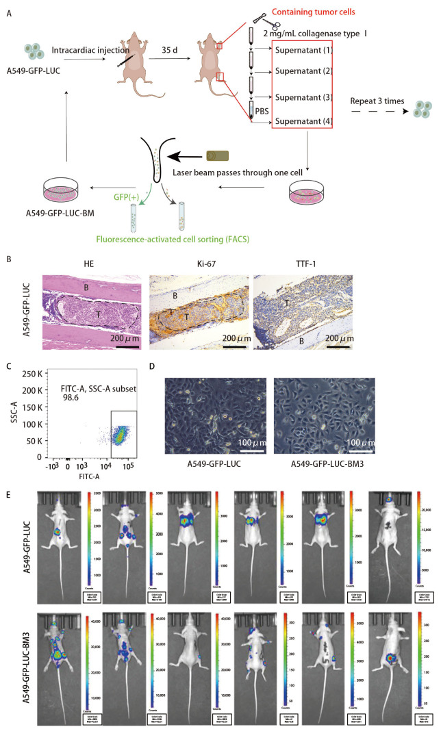
Figure 1. Establishment of the A549-GFP-LUC-BM3 bone metastasis model. (A) Schematic workflow of three-round in vivo selection via left ventricular injection, bone harvest, FACS sorting, and re-injection. (B) Representative BLI images showing progressive bone colonization across rounds. (C) H&E staining of decalcified bone sections confirming metastatic tumor foci in bone marrow. (D) GFP fluorescence and morphology of BM3 cells compared to parental line.
Enhanced Malignant Phenotype In Vitro
Comprehensive in vitro characterization revealed that A549-GFP-LUC-BM3 exhibits significantly enhanced malignant properties compared to the parental A549-GFP-LUC line, supporting its bone-metastatic potential:
(1) Proliferation: CCK-8 assays showed significantly higher proliferation rates in BM3 cells (P < 0.05). Colony formation assays confirmed dramatically increased clonogenic capacity (P < 0.0001), indicating enhanced long-term proliferative fitness.
(2) Migration and invasion: Scratch wound healing assays demonstrated accelerated lateral migration. Transwell migration and Matrigel invasion assays confirmed significantly enhanced motility (P < 0.01 to P < 0.001), consistent with the increased metastatic capacity observed in vivo.
(3) EMT marker expression: Western blot analysis revealed characteristic epithelial-mesenchymal transition (EMT) changes, including downregulation of E-cadherin and upregulation of N-cadherin and Vimentin, providing a molecular basis for the enhanced migratory and invasive phenotype.
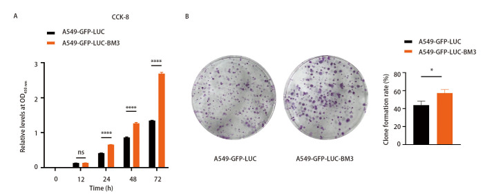
Figure 2. In vitro functional characterization of A549-GFP-LUC-BM3. (A) CCK-8 proliferation assay showing significantly higher growth rates in BM3 vs. parental cells. (B) Colony formation assay confirming increased clonogenic capacity. (C) Scratch wound healing assay demonstrating accelerated migration.
Our A549-Luc cells provide the quantitative sensitivity needed for rigorous preclinical metastasis research. Whether studying EMT, migration, or drug response, A549-Luc delivers reliable in vivo readouts.
Order Now: A549 Cell Line | A549-Luc Cell Line | A549-Luc-GFP Cell Line
EMT Reprogramming and In Vivo Metastatic Validation
The EMT phenotype observed in vitro directly translated to enhanced in vivo metastatic behavior. Western blot analysis confirmed that BM3 cells underwent a partial EMT, with reduced E-cadherin (epithelial marker) and increased N-cadherin and Vimentin (mesenchymal markers). This molecular reprogramming is consistent with the canonical EMT pathway that enables epithelial tumor cells to acquire migratory and invasive properties essential for extravasation and bone colonization.
In vivo validation experiments confirmed that BM3 cells exhibited significantly higher bone metastasis incidence and burden compared to parental A549-GFP-LUC cells when injected via the left cardiac ventricle. BLI monitoring enabled real-time, non-invasive tracking of metastatic progression, with signals progressively increasing in the spine, femur, and tibia over the 6–8 week observation period. Ex vivo GFP fluorescence of harvested bone tissues further confirmed the bone-tropic dissemination pattern.
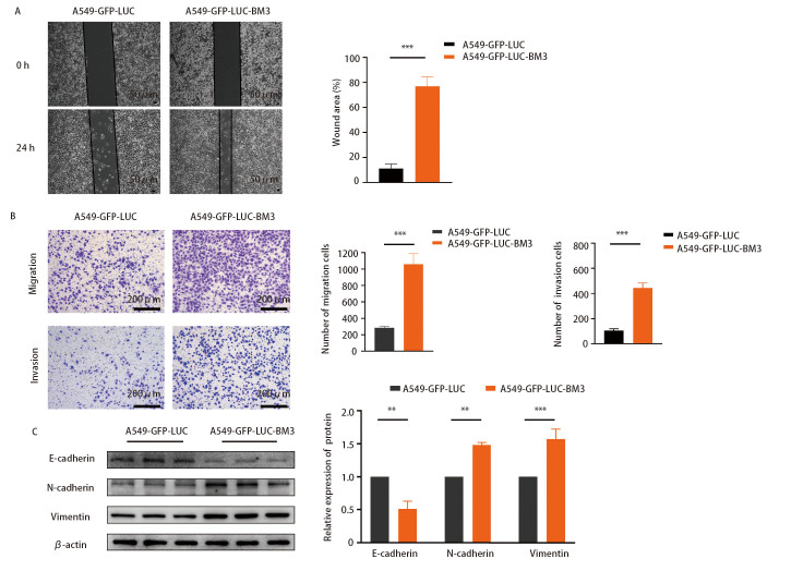
Figure 3. EMT marker expression and in vivo metastatic validation. (A) Western blot analysis of EMT markers (E-cadherin, N-cadherin, Vimentin) in BM3 vs. parental cells. (B) Scratch wound healing assay at 0h and 24h showing accelerated BM3 migration. (C) Transwell migration and Matrigel invasion assays confirming enhanced invasive capacity. (D) Quantitative analysis of migrated/invaded cells.
Transcriptomic Characterization of the Bone-Metastatic Phenotype
To elucidate the molecular mechanisms underlying the enhanced bone-metastatic potential, the investigators performed RNA-seq transcriptomic profiling comparing A549-GFP-LUC-BM3 to the parental line. The analysis identified 2,954 differentially expressed genes (DEGs), with 1,021 upregulated and 1,933 downregulated in the BM3 subline, revealing extensive transcriptional reprogramming associated with the metastatic phenotype.
Gene Ontology (GO) functional enrichment analysis highlighted cell adhesion, extracellular matrix organization, and signaling receptor activity as the most significantly enriched biological processes and molecular functions—consistent with the enhanced migratory and invasive behavior of BM3 cells. Cellular component analysis revealed enrichment in cell periphery, plasma membrane, and extracellular matrix terms.
KEGG pathway analysis identified several key pathways, including the NF-κB signaling pathway, cell adhesion molecule (CAM) pathways, and cytochrome P450-mediated drug metabolism pathways. The activation of NF-κB signaling is particularly noteworthy, as this pathway has been strongly implicated in bone metastasis progression across multiple cancer types, including lung adenocarcinoma.
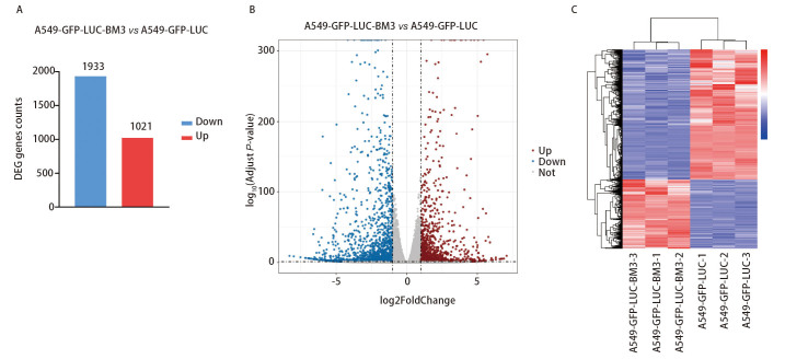
Figure 4. Transcriptomic analysis of A549-GFP-LUC-BM3. (A) Volcano plot showing differentially expressed genes between BM3 and parental cells (2,954 DEGs total). (B) Heatmap of top differentially expressed genes across samples. (C) Summary statistics of upregulated and downregulated gene counts.
Reliable in vivo tumor models are the foundation of impactful preclinical metastasis research. Our A549-Luc cell line offers stable dual-reporter expression, confirmed mycoplasma-free status, and proven performance in both subcutaneous and metastatic models.
Order Now: A549 Cell Line | A549-Luc Cell Line | A549-Luc-GFP Cell Line
Pathway Analysis and Therapeutic Implications
The comprehensive transcriptomic and functional data from this study provide a multi-layered understanding of the molecular drivers of lung adenocarcinoma bone metastasis. The enrichment of cell adhesion and extracellular matrix pathways in BM3 cells suggests that disruption of tumor-stroma interactions represents a promising therapeutic strategy for preventing or treating bone metastatic disease.
The identification of NF-κB signaling as a key activated pathway is particularly relevant for drug development. NF-κB inhibitors, including both small molecules and natural compounds, could be evaluated using the A549-GFP-LUC-BM3 model to assess their efficacy in blocking bone colonization and metastatic outgrowth. Similarly, the cytochrome P450 drug metabolism pathway enrichment may inform pharmacokinetic considerations for anti-metastatic therapies.
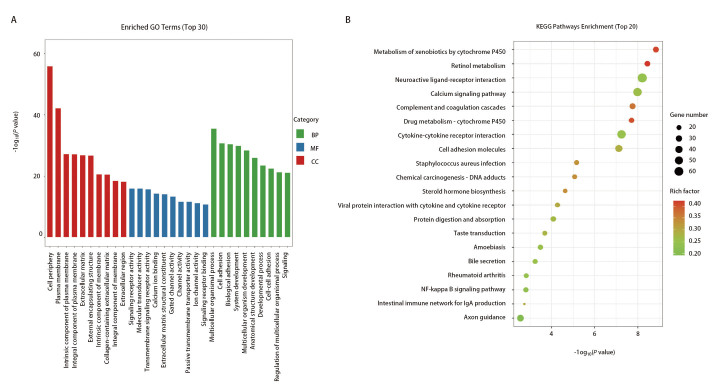
Figure 5. GO and KEGG pathway enrichment analysis. (A) GO biological process enrichment showing cell adhesion and extracellular matrix organization as top terms. (B) GO cellular component analysis. (C) GO molecular function enrichment. (D) KEGG pathway analysis highlighting NF-κB signaling, CAMs, and drug metabolism pathways.
Discussion
This study by Lu and colleagues establishes a powerful paradigm for lung adenocarcinoma bone metastasis research. The key innovations include: (1) a systematically selected high bone-metastatic cell line with dual-fluorescent reporters enabling both in vivo and ex vivo tracking; (2) comprehensive in vitro characterization confirming enhanced proliferation, migration, and EMT reprogramming; and (3) transcriptomic profiling revealing key molecular pathways driving the bone-metastatic phenotype.
The three-round in vivo selection approach ensures that the BM3 subline has undergone genuine Darwinian selection for bone-tropic fitness, rather than merely displaying an in vitro-adapted phenotype. The EMT reprogramming observed in BM3 cells is consistent with the well-established role of EMT in cancer metastasis, and the identification of NF-κB signaling as a key activated pathway provides a concrete molecular target for therapeutic intervention.
For the preclinical research community, this model addresses a critical gap in available tools for studying lung cancer bone metastasis. The dual-reporter design (luciferase for non-invasive BLI, GFP for histological validation) enables efficient longitudinal monitoring with reduced animal numbers, while the bone-metastatic phenotype provides a clinically relevant platform for evaluating anti-metastatic drug candidates.
Conclusion
The A549-GFP-LUC-BM3 cell line represents an indispensable tool for lung adenocarcinoma bone metastasis research and preclinical drug discovery. By combining dual luciferase/GFP reporters with three rounds of in vivo bone selection, the researchers have generated a model that faithfully recapitulates the molecular and phenotypic features of bone-metastatic disease. The Lu et al. study provides a methodological blueprint for establishing bone-metastatic models and identifies key pathways—including NF-κB signaling and cell adhesion molecule pathways—as promising therapeutic targets for preventing or treating lung cancer bone metastasis.
Our A549-Luc cell line provides the foundation for building bone-metastatic research models. Stable dual-reporter expression, well-characterized biology, and proven in vivo performance.
Order Now: A549 Cell Line | A549-Luc Cell Line | A549-Luc-GFP Cell Line
References
1. Lu Y, Qiu R, Deng Y, Liu X, Du Y. Establishment of dual fluorescent labeled human high bone metastasis lung adenocarcinoma cell line and transcriptomic characterization analysis. Zhongguo Fei Ai Za Zhi. 2024;27(4):257-265. doi:10.3779/j.issn.1009-3419.2024.101.09
