Cancer Cells Promote Immune Evasion in Tumor Liver Metastasis via VSIG4-CD5 Interaction with T Cells
Liver metastasis represents one of the most common forms of metastasis for various solid tumors. Once patients develop liver metastases, the prognosis often worsens significantly. Clinically, liver metastases show a considerably lower response rate to T cell immune checkpoint inhibitors compared to primary tumors or metastases in other organs. As the body's largest metabolic organ, the liver possesses intrinsic immune tolerance properties, which may be exploited by metastatic tumor cells to evade immune clearance. However, the molecular mechanisms underlying this process have long lacked systematic elucidation.Kupffer cells (KCs) are the most abundant tissue-resident macrophages in the liver, residing within the hepatic sinusoids. They possess the capacity to phagocytose tumor cells and present antigens, while also highly expressing various immunosuppressive molecules. Whether these cells play an anti-tumor or pro-tumor role in liver metastasis remained unclear until this study. This work systematically elucidates the molecular mechanism by which Kupffer cells regulate T cell responses and promote immune evasion in liver metastasis via the VSIG4-CD5 immune checkpoint pathway.
01. KCs have protumoral function by suppressing T cell responses
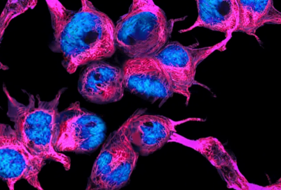
The study first identified the primary cell types responsible for phagocytosing tumor debris and presenting antigens in liver metastasis. Using splenic injection of ZsGreen-labeled B16F10 melanoma cells combined with intravital imaging, they found that resident F4/80⁺Tim-4⁺ Kupffer cells were the main cell type engulfing tumor debris. Using OVA as a model tumor antigen, they measured the levels of H-2Kᵇ-SINFEKL peptide-MHC I complexes on the surface of various hepatic antigen-presenting cells (APCs). Kupffer cells displayed significantly higher levels of antigen peptide-MHC I complexes on their surface compared to dendritic cells, B cells, monocyte-derived macrophages (MDMs), and liver sinusoidal endothelial cells (LSECs). Time-lapse intravital imaging further revealed long-lasting, high-frequency interactions between Kupffer cells and CD8⁺ T cells.To evaluate the functional impact of Kupffer cells on T cell responses, the researchers used Clec4f-iDTR mice to selectively deplete Kupffer cells. Results showed that upon Kupffer cell depletion, the number of CD8⁺ T cells and the proportion of CD62LˡᵒCD44⁺ effector cells significantly increased in the liver. In contrast, mice lacking cDC1s exhibited impaired CD8⁺ T cell responses and accelerated liver metastasis progression. This suggests that while cDC1s are crucial for T cell priming in secondary lymphoid organs to induce effective anti-tumor responses, Kupffer cells, despite their efficient cross-presentation capacity in the liver, negatively regulate CD8⁺ T cell responses.
Optimize your cancer immuno research with our OVA cell moldels.
Order now! >>B16F10-OVA Cell Line, B16F10-LUC-OVA Cell Line

02. KC-expressed VSIG4 has opposing effects on lowly antigenic versus highly antigenic LM
To identify key molecules mediating the negative regulatory function of Kupffer cells, the researchers generated Timd4⁻/⁻, Cd163⁻/⁻, and Vsig4⁻/⁻ knockout mice and examined their impact on B16F10 liver metastasis. Results showed that only Vsig4⁻/⁻ mice exhibited a significant phenotype of reduced liver metastasis. In an MC38 spontaneous liver metastasis model, Vsig4⁻/⁻ mice also showed a significantly lower incidence of metastasis. Subcutaneous tumor models revealed that VSIG4 knockout did not affect subcutaneous tumor growth, suggesting a liver-specific pro-tumor function for VSIG4.Tracking tumor growth dynamics via intravital imaging revealed no significant difference in tumor burden between Vsig4⁻/⁻ and wild-type mice on days 1 and 3 post-tumor inoculation, indicating that VSIG4 does not affect initial tumor seeding or the innate immune control phase. Differences emerged from days 7-10, when adaptive immunity begins to dominate tumor control. Flow cytometry analysis showed that Vsig4⁻/⁻ mice had increased numbers of CD8⁺ T cells in the liver, with enhanced activation, proliferation, and effector function, as well as increased tumor infiltration. CD8⁺ T cell depletion completely abolished the difference in tumor burden between wild-type and Vsig4⁻/⁻ mice, confirming that the effect of VSIG4 is T cell-dependent.
When using the highly immunogenic B16F10-OVA to establish a liver metastasis model, Vsig4⁻/⁻ mice paradoxically showed a higher liver metastatic burden than wild-type mice. KC-specific VSIG4 knockout mice similarly exhibited the phenotype of reduced B16F10 (low immunogenicity) but increased B16F10-OVA (high immunogenicity) metastasis. This indicates that VSIG4 has completely opposing effects on low versus high immunogenicity liver metastasis.

03. CD5 is a ligand for VSIG4
The ligand for VSIG4 on T cells has long been unknown. The researchers employed GST pull-down combined with liquid chromatography-mass spectrometry to identify VSIG4-binding proteins from lysates of anti-CD3/CD28-stimulated T cells. Among 19 candidate membrane proteins, CD5 attracted their greatest attention. Pull-down assays showed that VSIG4-GST could precipitate GFP-CD5; deleting the VSIG4 extracellular domain completely abolished binding, while deleting the intracellular or transmembrane domain did not affect binding. Domain mapping of CD5 revealed that the SRCR2 domain is critical for binding to VSIG4, with its deletion almost completely eliminating binding. ELISA confirmed a dose-dependent, direct binding between VSIG4-ECD and CD5-ECD, which is conserved between humans and mice. Complement components C3b and iC3b did not compete for binding, indicating that VSIG4-CD5 binding is complement-independent.Cellular binding assays showed that VSIG4-Fc specifically bound to NIH3T3 cells overexpressing CD5, and this binding could be completely abolished by CD5 knockout. On primary T cells, VSIG4-Fc specifically bound to activated CD8⁺ T cells, and this binding was strictly dependent on CD5 expression.

04. VSIG4–CD5 interaction inhibits T cell activation and activation-induced cell death in vitro
To elucidate the cellular signaling effects of VSIG4-CD5 engagement, the researchers performed in vitro T cell stimulation assays. Following anti-CD3/CD28 stimulation of wild-type mouse cells, VSIG4-Fc significantly inhibited T cell activation and apoptosis. The addition of a CD5 antibody partially reversed this inhibition. Furthermore, Cd5⁻/⁻ T cells were unaffected by VSIG4-Fc treatment in terms of activation.Mechanistically, VSIG4-Fc treatment enhanced the co-immunoprecipitation of CD5 with Cbl/Cbl-b, while concurrently reducing the phosphorylation levels of the TCR-proximal signaling molecules ZAP70 and LCK, as well as PLCγ1. This indicates that VSIG4 engagement promotes the recruitment of c-Cbl and Cbl-b to the CD5 signalosome, thereby inhibiting T cell activation and activation-induced cell death (AICD).

05. VSIG4–CD5 interaction bidirectionally regulates antitumor T cell responses
To validate the bidirectional regulatory function of the VSIG4-CD5 axis in vivo, the researchers employed an OT-I adoptive transfer system, transferring CD8⁺ T cells recognizing the OVA antigen into tumor-bearing mice. Using two OVA variants, G4 and N4, in a low-antigenicity B16F10-G4 tumor model, VSIG4 deficiency led to a significant increase in the number of OT-I cells in the liver, with elevated CD69 expression. However, cleaved caspase-3 levels, PD-1/Tim-3 double-positivity, and TOX expression showed no significant changes. This indicates that VSIG4 deficiency allows low-affinity T cells to gain enhanced activation without progressing to AICD or terminal exhaustion.In the high-antigenicity B16F10-N4 tumor model, VSIG4 deficiency resulted in decreased OT-I cell numbers, elevated CD69, but significantly increased cleaved caspase-3 and Fas, as well as increased PD-1/Tim-3 double-positivity and TOX expression. VSIG4 deficiency promoted the accumulation of Ly108⁺CD69⁺ precursor exhausted T cells, suggesting that VSIG4 normally protects high-affinity T cells from AICD and over-exhaustion. The phenotype of Cd5⁻/⁻ mice was consistent with that of Vsig4⁻/⁻. Vsig4⁻/⁻Cd5⁻/⁻ double knockout mice showed no significant difference in tumor burden compared to Cd5⁻/⁻ mice, demonstrating that the function of VSIG4 must be mediated through CD5.

06. VSIG4 shapes the immune landscape of metastatic liver tumors
To assess the impact of the VSIG4-CD5 axis at the tumor population level, the researchers co-injected high-immunogenicity MC38-OVA and low-immunogenicity MC38 at a 1:1 ratio. In wild-type mice, liver metastases predominantly originated from the low-immunogenicity MC38; however, in Vsig4⁻/⁻ mice, the proportion of high-immunogenicity MC38-OVA significantly increased. Single-cell RNA sequencing analysis revealed that in the livers of Vsig4⁻/⁻ mice, macrophages were decreased while T cell proportions were increased. Tumor-associated macrophages (TAMs) exhibited a pro-inflammatory signature, with upregulation of genes such as Cxcl1, Il1b, and Cxcl10. In T cells, effector/memory-related genes were upregulated, while exhaustion-related genes were downregulated. The transcriptomic changes in Kupffer cells from Vsig4⁻/⁻ mice were minimal, suggesting that VSIG4 primarily shapes the immune microenvironment indirectly by modulating T cells. TCR sequencing showed increased proportions of hyper-expanded and large clones among hepatic CD8⁺ T cells in Vsig4⁻/⁻ mice, along with higher clonal diversity. Staining with 6 MC38 neoantigen peptide-MHC tetramers revealed that Vsig4⁻/⁻ mice exhibited significantly enhanced specific CD8⁺ T cell responses to 5 of the neoantigens.

07. Blockade of VSIG4–CD5 interaction has therapeutic potential against LM
Finally, the researchers immunized alpacas with the mouse VSIG4 extracellular domain, constructed a phage display nanobody library, and screened 15 candidate anti-VSIG4 nanobodies, which were engineered as human IgG1 Fc fusion proteins. Among them, anti-VSIG4-5 displayed strong antigen-binding affinity and almost completely blocked the binding between VSIG4 and CD5.In established B16F10 liver metastasis models, anti-VSIG4-5 monotherapy only slightly inhibited tumor growth. B16F10 liver metastases are intrinsically resistant to anti-PD-L1 therapy. However, VSIG4 knockout rendered mice responsive to anti-PD-L1 treatment. Combination therapy with anti-VSIG4-5 and anti-PD-L1 produced a significant synergistic effect: tumor burden was substantially reduced, and the number, effector memory phenotype, and Granzyme B/IFN-γ expression of hepatic CD8⁺ T cells were significantly enhanced. Even when combination therapy was initiated on day 10 post-tumor inoculation (established metastases), it still significantly prolonged mouse survival.

Summary
This study elucidates the molecular mechanism by which Kupffer cells promote immune evasion in liver metastasis via the VSIG4-CD5 immune checkpoint pathway. Through the interaction of VSIG4 with CD5 on T cells, Kupffer cells achieve bidirectional regulation of T cell responses: suppressing T cell activation under low immunogenicity conditions, while protecting T cells from AICD under high immunogenicity conditions. The study also developed anti-VSIG4 nanobodies, demonstrating that combined therapy with anti-PD-L1 exerts synergistic effects in advanced liver metastasis models, providing a potential intervention strategy to overcome immunotherapy resistance in liver metastasis.
Reference:
Zhou X, Liu W, Hu J, et al. Kupffer cell calibration of T cell responses via VSIG4–CD5 interaction promotes tumor evasion. Nature Immunology (2026). doi: 10.1038/s41590-026-02510-w.
