Applications of C6 Cells in Neurotoxicology and Neuroprotection Research
Introduction
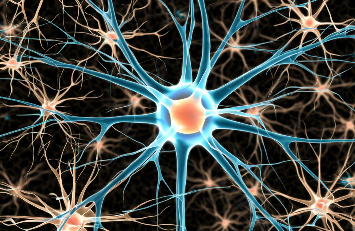
The health and function of the central nervous system depend not only on the proper functioning of neurons but also on the critical support provided by glial cells. Among glia, astrocytes act as essential support units, responsible for nutrient supply, ion homeostasis, and neuronal protection. When these support cells are damaged, neuronal function and survival are severely compromised. Therefore, evaluating the potential impact of exogenous substances on astrocytes is an important part of neuroscience research. The rat C6 glioma cell line has become a standardized in vitro tool for this field because it stably expresses astrocyte markers and is easy to culture.
Does your research require a reliable astrocyte model? Our C6 cells are the classic tool for studying neuron-glia interactions. Learn more>>
A Platform for Neurotoxicity Screening
During the early stages of drug development or environmental safety assessment, an efficient platform is needed to rapidly screen numerous compounds for potential neurotoxicity. C6 cells provide an ideal initial screening model due to their homogeneity and reproducibility. Researchers expose C6 cells to specific concentrations of test substances, such as new drug candidates, environmental pollutants (e.g., heavy metals like mercury and lead), or nanomaterials. The impact on the glial cells is then evaluated using a series of quantitative metrics.
Specific detection endpoints include:
Cell Viability: Assays such as MTT or CCK-8 are used to measure the metabolic activity of the cells, which directly reflects the cytotoxicity level of the compound.
Oxidative Stress: The production of intracellular reactive oxygen species (ROS) is measured using fluorescent probes like DCFH-DA to assess whether the compound induces oxidative damage.
Mitochondrial Function: Changes in the mitochondrial membrane potential are detected with probes like JC-1 to determine if the cell's "powerhouse" is compromised.
Inflammatory Response: The release or expression of inflammatory cytokines (e.g., Tumor Necrosis Factor-α, Interleukin-6) is quantified in the culture medium or within the cells using ELISA or qPCR.
Together, these data provide an initial profile of a compound's toxicity toward astrocytes, helping to eliminate substances with high neurotoxic risk at an early stage.
Neuron-Glia Co-culture Systems
A single C6 cell culture model cannot fully replicate the complex intercellular communication within the brain. Neuronal health is largely dependent on signals and support from astrocytes. Therefore, establishing neuron-glia co-culture systems is a key step toward investigating the indirect mechanisms of neurotoxicity.
In these systems, C6 cells are typically cultured together with primary hippocampal or cortical neurons. A common method involves the use of a Transwell insert system, where C6 cells are cultured in the bottom well and neurons are grown on a porous membrane in the top insert. This setup allows for chemical signaling between the two cell types through the shared medium but prevents physical contact. This model enables researchers to answer a core question: does a toxin kill neurons directly, or does it cause neuronal damage indirectly by first injuring C6 cells and impairing their protective functions, such as the secretion of neurotrophic factors? Differentiating between these mechanisms is instructive for understanding toxicity and developing targeted protective strategies.
Screening for Neuroprotective Drugs
Once reliable neurotoxicity models are established, the C6 cell line can also be used to screen and evaluate drugs with neuroprotective potential. The experimental design follows a "damage-then-protect" logic. Researchers first treat C6 cells with a known neurotoxin (e.g., glutamate, hydrogen peroxide, or MPP+) to induce a clear damage phenotype, such as elevated oxidative stress or reduced cell viability.
Subsequently, candidate neuroprotective drugs are co-incubated with the toxin or added after the toxin-induced damage. The protective effect of the drug is determined by measuring its ability to reverse or mitigate these damage indicators. For example, an effective neuroprotective agent might:
1. Increase the survival rate of C6 cells in the toxic environment.
2. Reduce the level of toxin-induced ROS.
3. Promote the secretion of additional neurotrophic factors from C6 cells (such as Brain-Derived Neurotrophic Factor, BDNF, or Glial Cell-Derived Neurotrophic Factor, GDNF), which can further protect neurons in a co-culture system.
Through this process, the C6 cell model offers a pathway for the rapid and efficient screening of drugs that can enhance the brain's own protective mechanisms.
Simulate the complex brain microenvironment in vitro. Our C6 cells are the ideal choice for building neuron-glia co-culture systems. Check now>>
Conclusion
In summary, the application of the C6 cell line has expanded well beyond its original scope as a tumor model. As a stable and easy-to-handle astrocyte-like model, it provides an efficient and economical in vitro platform for early-stage neurotoxicology screening, for studying complex neuron-glia interactions, and for the development of neuroprotective drugs. The proper utilization of this classic tool can provide valuable data to support research aimed at safeguarding central nervous system health.
References
[1]Benda, P., et al. (1968). Differentiated rat glial cell strain in tissue culture. Science, 161(3839), 370–371.
[2]Stelmashook, E. V., et al. (2019). Neuroprotective effect of erythropoietin-derived peptide in a co-culture of C6 glioma cells and hippocampal neurons under oxygen-glucose deprivation. Molecular Biology, 53(4), 609-618.
[3]Li, Q., et al. (2017). Protective effects of puerarin against lead-induced C6 glioma cell injury. Neural Regeneration Research, 12(1), 113-120.
[4]Foo, M., et al. (2021). The effects of silver nanoparticles on neuronal and glial cells. International Journal of Molecular Sciences, 22(13), 6982.
