The Role of Tumor-Antigen-Specific CD103+CD8+T Cells in Lung Metastasis of Breast Cancer: Insights from EO771-OVA Cells
1. Research Background
Immune checkpoint blockade therapy succeeds in cancer treatment by reactivating effector immune cells, but some patients have no response because their tumor microenvironment presents an "immune-cold" phenotype. Effector immune cell deployment (EICD) is a key part of immune surveillance; its defects lead to tumor immune escape. The long-range regulatory role and mechanism of adaptive immunity in distant metastasis have not been clarified. This study aims to clarify the role of tumor-antigen-specific CD103+CD8+T cells in breast cancer lung metastasis, define the molecular mechanisms of their initiation, recruitment and functional regulation, and reveal the impact of tumor microenvironment on the fate of these cells.
To achieve accurate experimental results in your breast cancer metastasis research, choose our high-purity EO771-OVA cells—contact us now to get your sample and accelerate your research progress! Order now>>
2. Research Design
2.1 Clinical Sample Analysis
Axillary lymph node samples from 533 newly diagnosed breast cancer patients were included. The distribution of CD103+CD8+T cell subsets was analyzed by immunofluorescence staining. Metastatic lesion samples from lung, liver, bone, brain and other sites were collected to analyze the correlation between cell infiltration and metastatic load.
2.2 Animal Models and Experiments
Mouse models of lung metastasis of breast cancer (EO771, 4T1) and melanoma (B16F10) were established. Tumor progression was tracked by luciferase or mCherry labeling. Conditional knockout mice (Cd8Cre/+ItgaeloxP/+DTR-GFP) were generated to specifically deplete CD103+CD8+T cells. Kaede photo-conversion mice and OT-I transgenic mice were used to verify cell origin and antigen specificity.
2.3 Molecular and Cellular Experiments
Single-cell RNA sequencing (scRNA-seq) was used to analyze immune cell subsets and signaling pathways in pre-metastatic lungs. Flow cytometry and immunofluorescence staining were used to detect cell phenotype, proliferation, apoptosis and cytokine secretion. Adoptive transfer experiments verified the functional specificity of CD103+CD8+T cells. EV isolation, purification and characterization were performed, and in vitro co-culture experiments verified their impact on AMs polarization. Inhibitors (1-MT, FTY720, etc.) were used to verify the role of targets such as IDO1 and CCL25/CCR9.
3. Research Results
3.1 CD103+CD8+T Cell Abundance in TDLN Correlates with Reduced Lung Metastasis
Analysis of CD8+T cell subsets in TDLN of 533 breast cancer patients showed that the abundance of CD103+CD8+T cells (resident memory phenotype) was significantly correlated with reduced risk of lung metastasis, while CD8+ central memory T cells (TCM) and effector memory T cells (TEM) had no such correlation. Higher abundance of these cells was associated with longer lung-specific metastasis-free survival in patients, which was applicable to all breast cancer subtypes. The infiltration of these cells in metastatic lung tissue was negatively correlated with metastatic load.
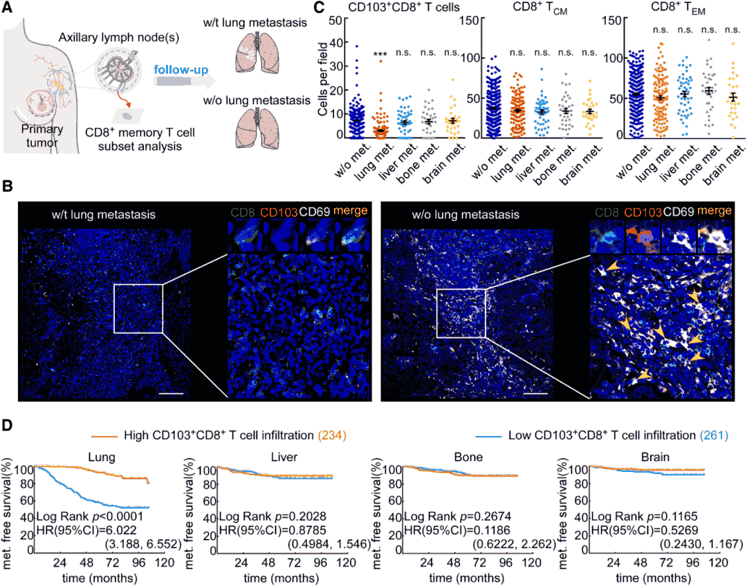
Fig1 Correlation between CD103+CD8+T cell deployment in TDLN and reduced lung metastasis
3.2 CD103+CD8+T Cells Accumulate in Non-Metastatic Lungs and Decrease Before Metastasis
In the EO771 breast cancer mouse model, CD103+CD8+T cells began to accumulate in lung tissue 2 weeks after tumor inoculation, reached a peak (51.2% of CD8+T cells) at 21-28 days, and then decreased sharply before the appearance of macroscopic metastasis. These cells were mainly located in lung tissue rather than blood vessels in pre-metastatic lungs, and showed enhanced proliferation ability, cytokine secretion (granzyme B, perforin, IFN-γ) and tumor-killing activity. Consistent results were obtained in 4T1 breast cancer and B16F10 melanoma models. In clinical samples, the infiltration of CD103+CD8+T cells in distant non-tumor lung tissue (more than 10cm from metastatic lesions) of patients with lung metastasis was significantly higher than that in paracancerous and metastatic tissues.

Fig2 CD103+CD8+T cells accumulate in non-metastatic lungs and decrease before metastasis
3.3 CD103+CD8+T Cells Inhibit Lung Metastasis Through Tumor-Antigen-Specific Cytotoxicity
Using conditional depletion mouse models, it was found that specific depletion of CD103+CD8+T cells significantly increased lung metastasis of breast cancer and melanoma and shortened mouse survival. Adoptive transfer of these cells (rather than TCM, TEM or effector T cells) significantly reduced lung metastatic load. Antigen-specific experiments showed that only CD103+CD8+T cells induced by EO771-OVA tumors could effectively recognize OVA257-264 peptides, specifically kill EO771-OVA cells and secrete IFN-γ, confirming that their role in inhibiting lung metastasis depends on tumor antigen specificity.
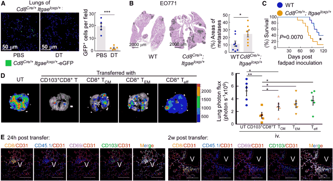
Fig3 CD103+CD8+T cells inhibit lung metastasis in vivo
3.4 Tumor-Specific CD103+CD8+T Cells Are Initiated in TDLN and Recruited to Lungs Before Metastasis
OT-I T cell tracking experiments showed that tumor-antigen-specific CD103+CD8+T cells first accumulated in TDLN (1 week after inoculation) and were detected in pre-metastatic lungs 3 weeks later. Kaede photo-conversion experiments confirmed that CD103+CD8+T cells infiltrating the lungs originated from TDLN rather than primary tumors or other lymph nodes. Dendritic cells (DC) in TDLN could effectively initiate CD103+CD8+T cells, and their initiating ability increased with the progression of metastasis. Inhibition of lymphocyte migration from TDLN using FTY720 significantly reduced the infiltration of CD103+CD8+T cells in the lungs, confirming that these cells need to circulate from TDLN to the lungs to function.
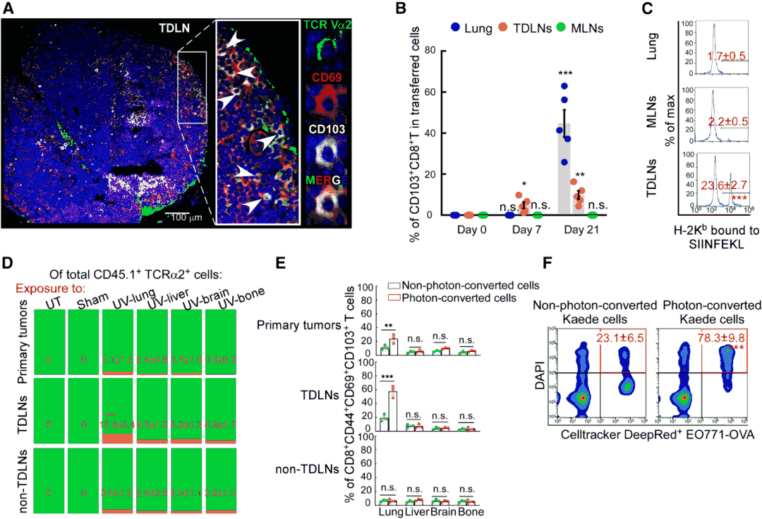
Fig4 Tumor-specific CD103+CD8+T cells are initiated in TDLN and recruited to lungs before metastasis
3.5 AMs Drive Lung CD103+CD8+T Cell Deployment Through CCL25/CCR9 and Induce Their Death Through IDO1
scRNA-seq analysis found that CD103+CD8+T cells specifically expressed CCR9, while its ligand CCL25 was mainly secreted by AMs. CCL25 secreted by AMs increased in the early stage of metastasis and decreased in the late stage, which was consistent with the infiltration dynamics of CD103+CD8+T cells. Neutralization of CCL25 or CCR9 blocked the recruitment of these cells to the lungs and accelerated metastasis. In the late stage of metastasis, AMs highly expressed IDO1, which reduced the function of CD103+CD8+T cells by inhibiting cytokine secretion and promoting apoptosis. Treatment with IDO1 inhibitor 1-MT could significantly increase the infiltration of these cells in the lungs, reduce metastasis and prolong survival, but had no effect in AMs-deficient Csf2-/- mice.
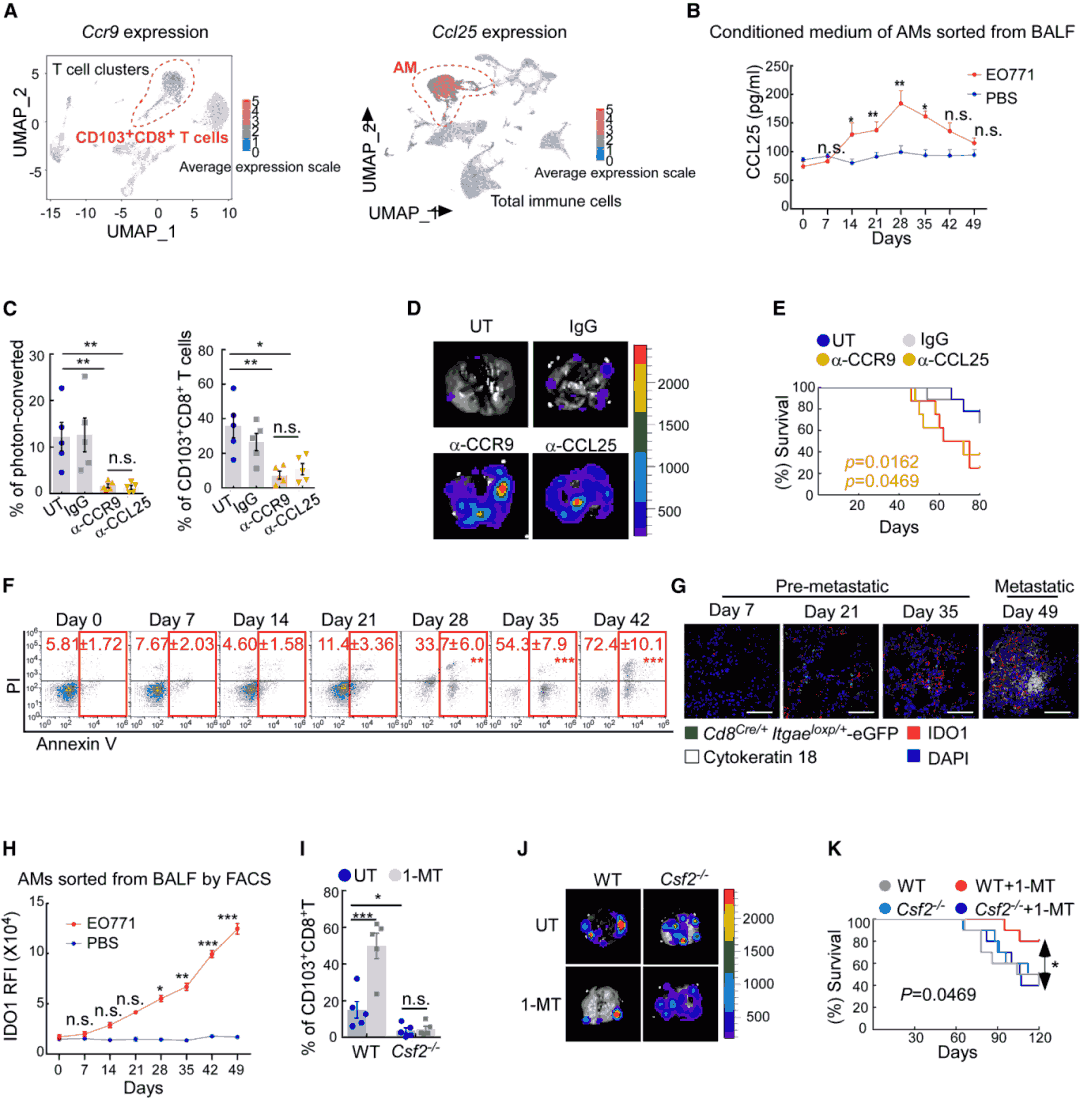
Fig5 AMs drive lung CD103+CD8+T cell deployment through CCL25/CCR9 and induce their death through IDO1
3.6 EVs Released by Tumors Differentially Polarize AMs at Different Stages
EVs released in the early and late stages of tumors (early-EV, late-EV) had no significant differences in morphology and diameter, but different cargo: early-EV were rich in proteins (such as TNF-α), while late-EV were rich in double-stranded DNA. Early-EV induced AMs to secrete CCL25 by activating the NF-κB pathway, and late-EV induced AMs to express IDO1 by activating the AIM2 pathway. In vivo experiments confirmed that injection of early-EV could increase the infiltration of CD103+CD8+T cells in the lungs and reduce metastasis, while late-EV had the opposite effect. This effect could be reversed by blocking ITGα6/ITGβ1 (lung-targeted uptake molecules) on the surface of EVs.
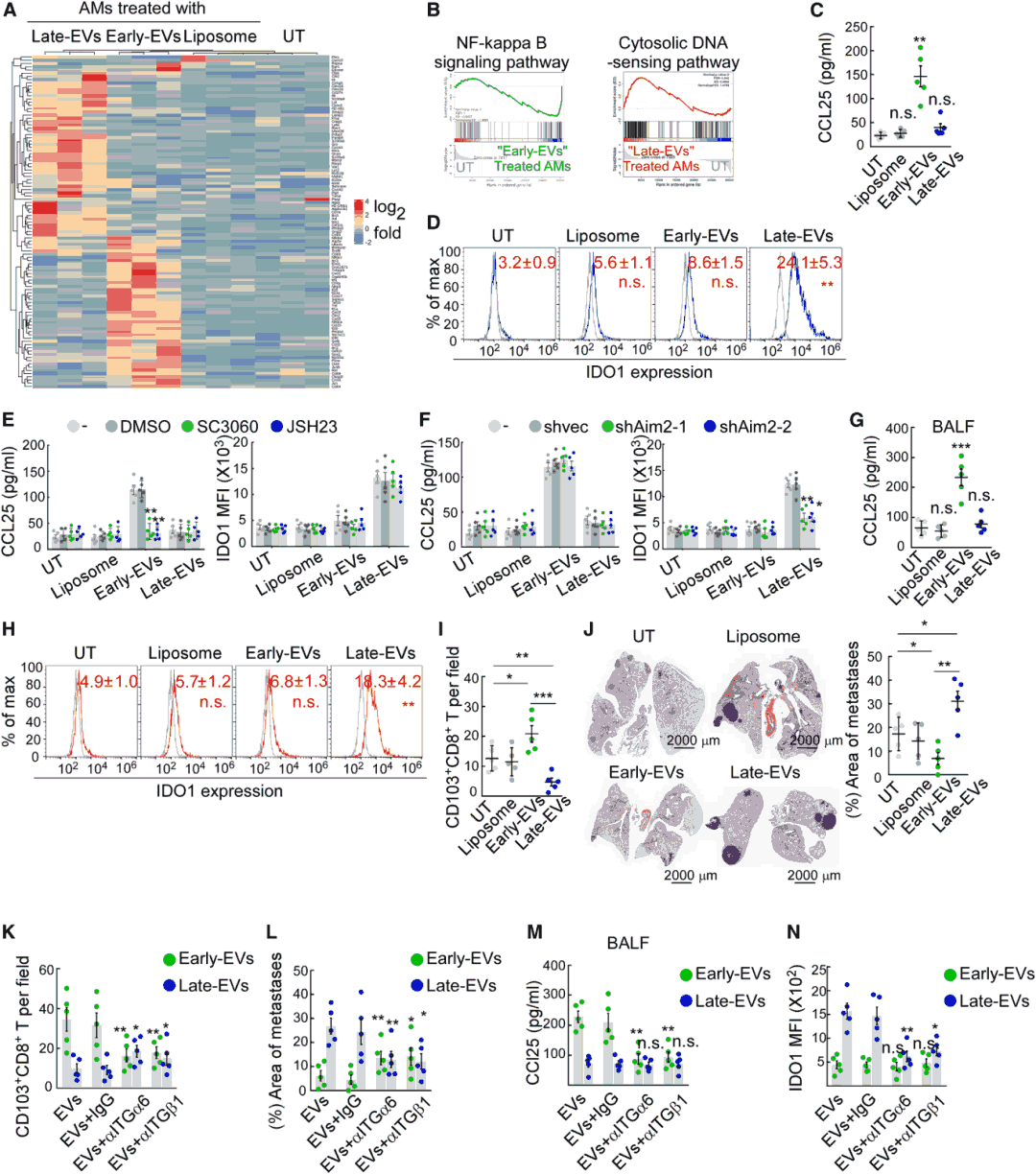
Fig6 EVs released by tumors differentially polarize AMs at different stages
Accelerate your cancer immunology research with our high-quality EO771-OVA cells, validated for stable antigen expression. Learn more>>
4. Results and Discussion
This study first reveals the long-range immune surveillance role of CD103+CD8+T cells in breast cancer lung metastasis, finds that tumor EVs polarize AMs through stage-specific cargo, dynamically regulate the recruitment and survival of CD103+CD8+T cells, and clarifies a new mechanism of pre-metastatic microenvironment remodeling.
The results show that after being initiated in tumor-draining lymph nodes, CD103+CD8+T cells are recruited to lung tissue through CCL25/CCR9 signals, and inhibit breast cancer lung metastasis through tumor-antigen-specific cytotoxicity. EVs released by tumors at different stages regulate the recruitment and survival of these cells by differentially polarizing alveolar macrophages. Targeting IDO1 or enhancing CCL25/CCR9-mediated cell recruitment can reconstruct EICD, providing a new therapeutic strategy for breast cancer lung metastasis.
References
Xing Y, Zhou Y, Wang R, et al. Long-range deployment of tumor-antigen-specific cytotoxic T lymphocytes inhibits lung metastasis of breast cancer. Dev Cell. Published online August 22, 2025. doi:10.1016/j.devcel.2025.08.003
