A Researcher's Guide to the GL261 Cell Line: All You Need to Know
Introduction
In the field of neuro-oncology, the selection of a preclinical model that faithfully mimics human disease while enabling the study of interactions with a competent immune system is paramount. The GL261 cell line stands out as such a model. As one of the most extensively used murine cell lines for studying glioblastoma (GBM), the most aggressive primary brain tumor, GL261 offers an unparalleled platform for exploring the tumor microenvironment and testing novel therapeutics, particularly immunotherapies. For researchers new to the field, a thorough understanding of GL261's background, characteristics, and applications is the first step toward designing rigorous and impactful experiments. This guide provides a systematic overview of the core knowledge required to master this powerful research tool.
A deep understanding of GL261's biological properties
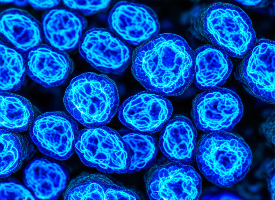
Morphology: Microscopically, GL261 cells typically exhibit a pleomorphic, glial- or astrocytic-like appearance, with some cells displaying spindle-shaped processes. They grow as an adherent monolayer in culture flasks.
Growth Properties: GL261 is a relatively fast-proliferating cell line. Its doubling time is generally reported to be between 24 and 35 hours, though this can vary slightly depending on culture conditions such as media formulation and serum concentration. Maintaining the cells in their logarithmic growth phase is critical for achieving consistent results in downstream applications, especially for in vivo tumor implantation.
Immunophenotype: This is one of the most valued attributes of GL261. As an immunogenic tumor, it can be recognized by the host's immune system. Key aspects of its immune profile include:
MHC Class I Expression: GL261 cells express Major Histocompatibility Complex (MHC) class I molecules on their surface. This is a prerequisite for the presentation of tumor antigens to cytotoxic T lymphocytes (CTLs), enabling immune-mediated attack.
PD-L1 Expression: The cells also express Programmed Death-Ligand 1 (PD-L1), an immune checkpoint molecule that tumors exploit to suppress T-cell activity. This feature makes the GL261 model an ideal platform for testing checkpoint inhibitors targeting the PD-1/PD-L1 axis.
Tired of inconsistent results? We offer rigorously validated GL261 line, ensuring your research data is highly reproducible. Click to order>>
GL261 holds an indispensable position in brain tumor research
The Gold Standard for Immunotherapy Research: As mentioned, its syngeneic origin and immunogenic nature make it the preeminent model for immuno-oncology studies. Researchers leverage it to assess checkpoint inhibitors, therapeutic vaccines, CAR-T cell therapies, and other strategies designed to mobilize the host immune system against the tumor.
High Fidelity to Human GBM Pathology: When GL261 cells are implanted orthotopically into the brains of C57BL/6 mice, the resulting tumors share striking histopathological similarities with human GBM. These features include:
High Invasiveness: Tumor cells diffusely infiltrate surrounding healthy brain tissue, creating an indistinct tumor margin.
Robust Angiogenesis: The tumors develop a rich, abnormal vascular network to sustain their growth.
Necrosis and Pseudopalisading: Central necrotic regions surrounded by hypercellular zones of tumor cells are often observed, which are classic hallmarks of human GBM.
Richly Characterized and Standardized: Decades of extensive use have resulted in a wealth of published data and standardized protocols for the GL261 model. This provides a robust foundation for new investigations and facilitates the comparison of results between different laboratories, thereby enhancing the trustworthiness and reliability of research findings.
Need a preclinical model that closely mimics human GBM pathology? Our GL261 cells are your ideal choice for generating trusted in vivo data. Order now>>
Conclusion
In summary, the GL261 cell line is far more than just a vial of cancer cells; it is a well-validated and highly characterized scientific instrument. Its syngeneic murine origin, immunogenic profile, and ability to faithfully recapitulate key pathological features of human GBM in vivo secure its irreplaceable role in fundamental brain tumor research and, most critically, in the preclinical evaluation of immunotherapies. Mastering its origins, biological properties, and applications is an essential prerequisite for any researcher dedicated to conquering glioblastoma.
References
[1] Ausman, J. I., Shapiro, W. R., & Rall, D. P. (1970). Studies on the chemotherapy of experimental brain tumors: development of an experimental model. Cancer Research, 30(9), 2394–2400.
[2] Szatmari, T., Lumniczky, K., Desaknai, S., Trajcevski, S., Hidy, D., Gyorgy, A., & Safrany, G. (2006). Detailed characterization of the mouse glioma 261 tumor model for experimental glioblastoma therapy. Cancer Science, 97(6), 546–553.
[3] Wainwright, D. A., Chang, A. L., Dey, M., Balyasnikova, I. V., Kim, C. K., Tobias, A., ... & Lesniak, M. S. (2014). Durable therapeutic efficacy utilizing combinatorial blockade against IDO, CTLA-4, and PD-L1 in mice with established brain tumors. Clinical Cancer Research, 20(20), 5290–5301.
[4] Sampson, J. H., Gunn, M. D., Fecci, P. E., & Ashley, D. M. (2020). Brain immunology and immunotherapy in brain tumours. Nature Reviews Cancer, 20(1), 12–25.
[5] Candolfi, M., Curtin, J. F., Nichols, W. S., Muhammad, A. G., King, G. D., Jaita, G., ... & Lowenstein, P. R. (2007). Intracranial glioblastoma models in preclinical neuro-oncology: neuropathological characterization and tumor progression. Journal of Neuro-oncology, 85(2), 133–148.
