
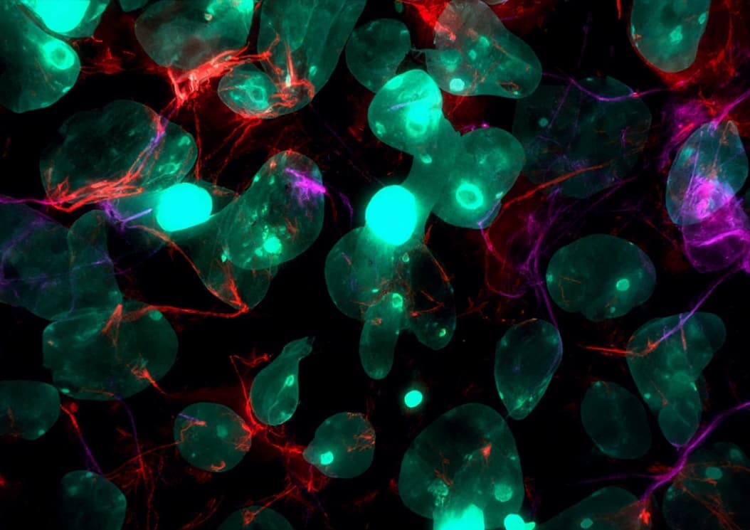
HEK-293T | A Novel Degrader Design Platform That Indirectly Recruits VHL via RIPK1, Breaking the Bottleneck of E3 Ligand
Published Online: July 8, 2026Source: Journal of Medicinal Chemistry
Research Team: Shen Yudao / Zhu Mingyan / Zhao Bo, Shanghai Jiao Tong University
Introduction
PROTAC (Proteolysis-Targeting Chimera) technology offers a novel avenue for innovative drug development. Since its conceptualization in 2001, over 20 molecules have entered clinical trials targeting various malignancies. Compared to traditional small-molecule inhibitors, PROTACs can degrade target proteins with high specificity, overcome drug resistance at low doses through catalytic action, and target "undruggable" proteins. However, their development is constrained by the scarcity of E3 ligase ligands—while human cells contain over 600 E3 ligases, only CRBN and VHL ligands are widely used in PROTAC design. PROTACs based on these two ligands have significant limitations: CRBN-based PROTACs may induce off-target degradation of neosubstrates such as IKZF1/3 and GSPT1; long-term use of CRBN- or VHL-based PROTACs can lead to resistance, for instance OVCAR8 cells developed resistance after four months of BET-PROTAC treatment. These drawbacks have driven exploration of novel E3 ligands and indirect recruitment strategies, including direct use of E3 ligands such as MDM2, IAPs, RNFs, DCAFs, and KEAP1, as well as strategies that indirectly recruit E3 by hijacking protein complexes (e.g., L3MBTL3/DCAF5, HSP90/E3s, GLP/SPOP). Building on this foundation, this study proposes the RIPK1-Mediated Targeting Chimera (RIMTAC) platform, which utilizes RIPK1 inhibitors to hijack the endogenous RIPK1-VHL complex for indirect VHL recruitment. Proof-of-concept validation was performed targeting BRD4, AKT, and JAK1, with mechanistic studies demonstrating that degradation activity depends on the RIPK1-VHL complex and operates through the ubiquitin-proteasome system.Results and Discussion
1. RIPK1 Inhibitors Do Not Disrupt the RIPK1-VHL Complex
To develop PROTACs capable of indirectly recruiting VHL, the researchers systematically reviewed VHL-binding proteins and found that while most are VHL substrates negatively regulated by VHL at the protein level, RIPK1, a key regulator of apoptosis, necroptosis, and inflammation, forms a complex with VHL under normoxic or mild hypoxic conditions. Notably, VHL does not affect RIPK1 protein stability but effectively inhibits its kinase activity. Based on this property, the researchers hypothesized that RIPK1 ligands could utilize the RIPK1/VHL complex as an indirect recruiter of VHL in PROTAC design.Using the RIPK1 inhibitor RIPK1i as a starting point, docking predictions identified its benzothiazole group as a solvent-exposed region, leading to synthesis of the tool compound Biotin-RIPK1i (biotin linked via a 4-unit PEG chain at this site) to verify whether RIPK1i binding interferes with RIPK1-VHL interaction. Biotin-RIPK1i successfully pulled down endogenous RIPK1 and VHL, indicating that RIPK1 inhibition does not disrupt this complex; co-IP experiments further confirmed their direct interaction. The RIPK1-VHL interaction is known to depend on EGLN1-mediated hydroxylation of RIPK1 Pro195 under normoxic conditions, and treatment with the EGLN1 inhibitor FG-4592 partially disrupted this interaction, consistent with previous reports. Collectively, these findings support the suitability of the RIPK1 inhibitor RIPK1i for designing RIMTACs through indirect VHL recruitment.

2. Design and Evaluation of BRD4 RIMTACs
Based on the above validation, the researchers designed RIMTACs using the RIPK1 inhibitor and selected BRD4 as a target protein to validate the strategy. By conjugating RIPK1i with the BRD4 inhibitor JQ1 via alkyl or PEG linkers of varying lengths, a series of 12 candidate BRD4 RIMTAC molecules were designed and synthesized, then evaluated for their ability to induce endogenous BRD4 degradation in HEK-293T cells. Compound 10, containing a 2-unit PEG chain, demonstrated the most potent BRD4 degradation activity at both 100 nM and 500 nM concentrations after 24 hours of treatment.
3. Compound 10 Induces BRD4 Degradation in HEK-293T Cells
In time-dependent degradation studies, compound 10 achieved 50% BRD4 degradation at 500 nM within 12 hours, reaching maximum degradation at 36 hours. Further studies showed concentration-dependent degradation, with a DC₅₀ of 54.12 nM and Dₘₐₓ of 83.3% after 24-hour exposure. To verify degradation of overexpressed BRD4, a BRD4-FLAG plasmid was constructed and transfected into HEK-293T cells; results showed that FLAG-tagged BRD4 was also degraded by compound 10 in a concentration-dependent manner. Whole-proteome mass spectrometry analysis assessing degradation selectivity showed that compound 10 maximally reduced BRD4 protein abundance in the proteome, while showing selectivity over other BET family proteins (BRD1, BRD2, BRD3, and BRD7), demonstrating that targeted protein degradation selectivity can be achieved using RIPK1 ligands to indirectly recruit VHL E3 ligase.
4. Compound 10-Mediated BRD4 Degradation Is Dependent on the Ubiquitin-Proteasome System
To elucidate the molecular mechanism, the researchers first investigated the potential role of the ubiquitin-proteasome system (UPS). Co-treatment with the proteasome inhibitor MG132 (5 μM) effectively blocked BRD4 depletion, strongly suggesting the proteasome is the ultimate executor. Further treatment with the NEDD8-activating enzyme inhibitor MLN4924 (1 μM), which is critical for Cullin-RING ligase (CRL) activation, significantly restored BRD4 levels, confirming dependence not only on the proteasome but also on functional CRL activity. Immunoblotting with anti-ubiquitin antibodies detected clear polyubiquitination signals, providing further evidence for UPS-mediated ubiquitination involvement. qRT-PCR confirmed that compound 10 did not alter BRD4 mRNA levels, ruling out transcriptional downregulation and confirming post-translational-level loss of BRD4. Additionally, washout experiments showed that BRD4 protein levels returned to baseline within 36 hours after compound removal, confirming the degradation effect is reversible and does not cause persistent disruption of endogenous BRD4 synthesis. These data collectively indicate that compound 10 acts as a catalytic degrader eliminating BRD4 protein through the ubiquitin-proteasome system.
5. Compound 10-Mediated BRD4 Degradation Requires Formation of a Quaternary Complex
As a RIMTAC molecule, compound 10 is designed to bridge BRD4 with RIPK1, the substrate receptor of the VHL E3 ligase complex, and its efficacy depends on simultaneous binding to both BRD4 and RIPK1. Competitive binding experiments showed that co-treatment with excess BRD4 inhibitor JQ1 (2 μM) or excess RIPK1 inhibitor RIPK1i (5 μM) along with compound 10 (500 nM) abolished BRD4 degradation, supporting a model in which both the warhead (BRD4-binding) and ligand (RIPK1-binding) moieties of the RIMTAC are indispensable—saturation of either binding site disrupts formation of the effective ternary complex (RIPK1-compound 10-BRD4). Further validation of whether BRD4 degradation requires functional integrity of the RIPK1-VHL complex: shRNA-mediated knockdown of either RIPK1 or VHL completely abolished compound 10-induced BRD4 degradation, confirming both are essential. The VHL inhibitor VH298, which disrupts VHL-HIF-1α interaction, was hypothesized to also disrupt VHL-RIPK1 interaction and thereby inhibit BRD4 degradation; experiments confirmed VH298 completely restored BRD4 levels in the presence of compound 10. Additionally, EGLN1-mediated hydroxylation of RIPK1 Pro195 is required for VHL binding under normoxic conditions; inhibiting this hydroxylation should disrupt VHL-RIPK1 interaction and eliminate BRD4 degradation. Treatment with the selective EGLN1 inhibitor FG-4592 indeed dissociated RIPK1 from VHL and abolished BRD4 degradation in the presence of compound 10, demonstrating that the degradation mechanism depends not merely on the presence of RIPK1 and VHL, but critically on their physiological interaction. Finally, to confirm formation of the required quaternary complex (BRD4-compound 10-RIPK1-VHL) for this RIMTAC system, co-IP experiments were performed, showing that both RIPK1 and VHL were effectively co-immunoprecipitated by FLAG-BRD4 in the presence of compound 10, consistent with the hypothesis.
6. Compound 10 Induces BRD4 Degradation in RAW264.7 Cells
The researchers further extended the degradation efficacy of compound 10 to other cellular contexts, selecting the murine macrophage cell line RAW264.7, widely used in studies of immune function, inflammatory responses, and cancer biology, as an example. Consistent with observations in HEK-293T cells, compound 10 effectively induced endogenous BRD4 degradation in a concentration-dependent manner, with a DC₅₀ of 179.1 nM and Dₘₐₓ of 65.63% after 24-hour treatment; the degradation was also time-dependent, with BRD4 levels reduced by 50% within 12 hours at 500 nM. Washout experiments showed gradual recovery of BRD4 protein levels to baseline within 24 hours after compound removal, indicating reversibility of degradation. Mechanistic exploration showed that competitive co-treatment with excess RIPK1 inhibitor RIPK1i or BRD4 ligand JQ1 along with compound 10 restored BRD4 protein levels, indicating degradation depends on formation of a ternary complex involving RIPK1, compound 10, and BRD4; furthermore, co-treatment with proteasome inhibitor MG132 or neddylation inhibitor MLN4924 completely abolished BRD4 degradation. qRT-PCR confirmed that compound 10 did not affect BRD4 mRNA levels, ruling out transcriptional downregulation. These results confirm that the BRD4 degradation activity of compound 10 is not limited to HEK-293T cells but is equally effective in RAW264.7 macrophages, with the same mechanism of degradation in both cell types.
7. Design and Evaluation of AKT RIMTACs
The researchers then attempted to extend the RIMTAC design strategy to a broader range of protein targets, selecting the kinase AKT as a study subject. Following established methodology, a series of candidate AKT-targeting RIMTAC molecules were designed and synthesized by conjugating the RIPK1 inhibitor RIPK1i with the known AKT inhibitor AKTi (AZD5363) via flexible PEG linkers of varying lengths (1-5 units). Systematic evaluation of these candidates for AKT degradation activity identified compound 14, which induced significant AKT depletion at 5 μM after 24-hour treatment. Further quantitative analysis showed compound 14 promoted AKT degradation in a concentration-dependent manner, with a DC₅₀ of 3.52 μM and Dₘₐₓ of 54.5% after 24 hours. In addition to concentration dependence, the kinetic profile of AKT degradation was examined: consistent with the kinetics of compound 10-mediated BRD4 degradation, compound 14 also induced time-dependent AKT loss, achieving approximately 50% degradation at 5 μM within 18 hours and maximum degradation at 48 hours. Mechanistically, pretreatment with the proteasome inhibitor MG132 or neddylation inhibitor MLN4924 significantly restored AKT protein levels, confirming UPS dependence; excess AKT inhibitor AKTi or RIPK1 inhibitor RIPK1i competitively inhibited AKT degradation, indicating formation of a ternary complex involving RIPK1, compound 14, and AKT; moreover, VH298 disruption of VHL-RIPK1 interaction rescued AKT levels in the presence of compound 14. Importantly, compound 14 did not alter AKT mRNA levels, ruling out transcriptional downregulation. These findings collectively validate successful extension of the RIMTAC design strategy to the AKT target.
8. Design and Evaluation of JAK1 RIMTACs
The researchers further investigated whether the RIMTAC strategy could be extended to the kinase JAK1, a known inflammatory driver. By conjugating the RIPK1 inhibitor RIPK1i with the known JAK1 inhibitor JAK1i (momelotinib) via linkers of varying lengths (specifically 1-7 unit alkyl chains and 1-5 unit PEG chains), 12 candidate JAK1 RIMTAC molecules were designed and synthesized. Evaluation of their ability to induce endogenous JAK1 degradation in HEK-293T cells at 500 nM for 24 hours showed that several molecules, including compounds 23, 24, 30, and 31, induced over 50% JAK1 degradation. Compound 24 was selected as the most potent JAK1 degrader for further characterization. Consistent with the previously described BRD4 and AKT degraders, compound 24 promoted JAK1 loss in a concentration- and time-dependent manner: concentration-dependent degradation showed a DC₅₀ of 322.8 nM, with Dₘₐₓ determined after 24-hour treatment; in time-dependent experiments, degradation was observed within 6 hours at 500 nM, with increasing degradation over time. This degradation was blocked by co-treatment with the proteasome inhibitor MG132 (2 μM) or neddylation inhibitor MLN4924 (1 μM), confirming UPS dependence; excess JAK1 inhibitor JAK1i (1 μM) or RIPK1 inhibitor RIPK1i (5 μM) competitively inhibited JAK1 degradation, supporting a mechanism involving formation of a ternary complex with RIPK1, JAK1, and compound 24; the VHL inhibitor VH298 (50 μM) also rescued compound 24-induced JAK1 degradation. Furthermore, compound 24 did not affect JAK1 mRNA levels, indicating post-translational regulation. These findings further validate the generality of the RIMTAC platform for rational design of degraders targeting multiple kinase targets.

Looking for HEK-293T cell lines for your medical studies? Visit our 293T series products>>293T cell line | HEK293T-EGFP | HEK293T-Luc | 293T-LUC-GFP
Summary and Outlook
Among the over 600 E3 ligases in nature, only a small number are targeted by small-molecule ligands for PROTAC design, with CRBN and VHL ligands remaining dominant. This study utilized a RIPK1 inhibitor to indirectly recruit the VHL E3 ligase complex, successfully designing and characterizing RIMTAC molecules targeting BRD4, AKT, and JAK1, with mechanisms confirmed to operate through UPS-mediated degradation. The poor oral bioavailability of traditional VHL ligands limits clinical translation, whereas RIPK1 inhibitors possess favorable pharmacokinetic properties, and the RIMTAC strategy holds promise to address this bottleneck. Simultaneously, the anti-inflammatory effect of RIPK1 inhibition itself, combined with degradation of inflammatory mediators such as BRD4 and JAK1, offers potential for synergistic therapeutic benefit. It should be noted that the oxygen sensitivity of the RIPK1-VHL interaction may limit application in severely hypoxic environments such as solid tumors. Overall, RIMTAC provides a new paradigm for indirect E3 ligase recruitment, representing a versatile platform for expanding the VHL toolbox with promising applications for degradation of inflammation-related proteins.For comprehensive details, please refer to the original publication:
https://doi.org/10.1021/acs.jmedchem.6c00897
