HT-1080 Cell Line Characteristics and Design Principles of Dual-Labeled Models
Introduction
The HT-1080 cell line, derived from a fibrosarcoma in a 35-year-old male, is a common model in cell biology. This cell line possesses a near-diploid karyotype and carries an activated N-ras oncogene. Due to its short growth cycle and ease of genetic manipulation, HT-1080 is an ideal tool for studying tumor proliferation, invasion, and drug responses. To achieve precise real-time monitoring, researchers integrated firefly luciferase (Luciferase) and green fluorescent protein (GFP) into these cells using genetic engineering. This dual-labeling system combines macro-dynamic observation with micro-histological analysis, significantly enhancing the multidimensionality of experimental data.
Origins and Core Characteristics of HT-1080 Cells
The HT-1080 cell line was established in 1974 from human fibrosarcoma tissue. In laboratory culture, these cells exhibit clear adherent growth patterns. Their genomic structure remains relatively stable, providing a foundation for complex gene editing.
Research data show that HT-1080 cells possess strong migratory capabilities. This trait is closely linked to the high levels of matrix metalloproteinases (MMPs) they express. Unlike common epithelial-derived tumor cells, HT-1080 retains the morphological and functional features of mesenchymal cells. This characteristic makes the line irreplaceable for studying sarcoma pathogenesis and developing targeted therapies. Furthermore, these cells demonstrate high tolerance for viral transduction, ensuring stable expression of exogenous genes.
Improve the efficiency of your screening assays by choosing the HT-1080 cell line as your reliable in vitro evaluation model. Consult us for purchasing options>>
Logic of Dual-Labeling: Combining Luc and GFP
To overcome the limitations of traditional experiments in quantifying tumor burden in real-time, researchers designed a dual-signal system containing Luc and GFP.
The primary purpose of Luciferase labeling is to enable bioluminescence imaging (BLI). Upon adding the substrate luciferin, the cells produce light signals invisible to the eye but detectable by instruments. The intensity of this signal correlates linearly with the number of viable cells. This allows researchers to monitor tumor growth dynamics within living animals continuously without sacrificing them.
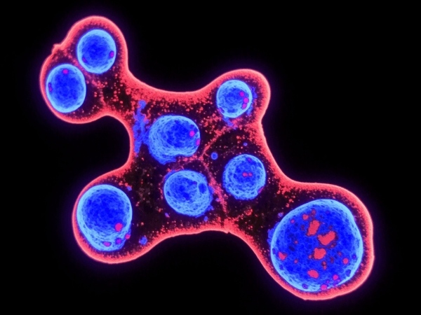
The introduction of Green Fluorescent Protein (GFP) focuses on spatial localization. GFP requires no substrate for excitation, making it suitable for confocal microscopy. Through GFP signals, researchers can observe the detailed distribution of HT-1080 cells within tissues and even capture single cells migrating near blood vessels. This dual-labeling design covers detection needs from the systemic level down to the single-cell level using two distinct optical signals.
Structural Advantages and Value of HT-1080-luc-gfp
The advantage of the HT-1080-luc-gfp model lies in the synergy of its signals. Regarding stability, stable cell lines generated via lentiviral transduction integrate Luc and GFP genes into chromosomes, ensuring signals are not lost during cell division.
In experimental logic, this model eliminates the data bias caused by single labeling. For instance, during anti-tumor drug screening, the Luc signal provides an overall efficacy assessment, while the GFP signal explains whether the drug altered cell morphology or distribution. Additionally, HT-1080-luc-gfp performs excellently in simulating tumor metastasis. Due to low background noise, researchers can detect tiny metastatic lesions in lungs or bone marrow, which is critical for evaluating the efficacy of drugs in preventing metastasis.
Choose HT-1080-luc-gfp cell to achieve precise quantitative monitoring of tumor growth via bioluminescent signals. Apply online now>>
Conclusion and Future Applications
The HT-1080-luc-gfp cell line not only retains the invasive traits of the original line but also achieves visualization and quantification through dual-labeling technology. This model simplifies experimental workflows and reduces data error. As in vivo imaging technology advances, this cell line will play a central role in evaluating novel anti-tumor therapies.
References
[1] Rasheed, S., et al. (1974). Characterization of a newly derived human sarcoma cell line (HT-1080). Journal of the National Cancer Institute.
[2] Brown, R., et al. (1984). Mechanism of activation of an N-ras gene in the human fibrosarcoma cell line HT1080. EMBO Journal.
[3] Contag, C. H., & Bachmann, M. H. (2002). Advances in in vivo bioluminescence imaging of gene expression. Annual Review of Biomedical Engineering.
[4] Chalfie, M., et al. (1994). Green fluorescent protein as a marker for gene expression. Science.
