Building Immunocompetent Models: Application of KLN205-Luc-GFP in Lung Cancer Immunotherapy
Introduction
Lung Squamous Cell Carcinoma (LUSC) accounts for approximately 25-30% of non-small cell lung cancer cases, exhibiting distinct pathogenic mechanisms and immune microenvironments compared to lung adenocarcinoma. However, most preclinical studies rely heavily on C57BL/6-background adenocarcinoma models (e.g., LLC1), leading to a lack of specificity in immunotherapy data for LUSC.
The KLN205-Luc-GFP cell line, a lung SCC line derived from DBA/2 mice, fills this gap by integrating a dual-reporter system. As a rare syngeneic model for lung SCC, it not only represents a specific pathological subtype but also serves as a precision tool for evaluating PD-1/PD-L1 inhibitors. The addition of Green Fluorescent Protein (GFP) alongside Luciferase (Luc) allows for a seamless transition from non-invasive whole-body imaging to single-cell level analysis.
The KLN205-Luc-GFP cell line features stable dual-reporter expression, enabling both longitudinal quantification of tumor burden and high-resolution histological tracking. View Product Details>>
Model Principle: Dual-Reporter Syngeneic Transplants
The primary principle of constructing immuno-oncology models is "immune matching." The KLN205-Luc-GFP cell line retains the original murine MHC Class I molecular characteristics (H-2d haplotype). When transplanted into immunocompetent DBA/2 mice, the tumor cells establish dynamic interactions with the host immune system, authentically recapitulating the "immuno-editing" process.
The Dual-Reporter Advantage:
Luciferase (Luc): Provides real-time, non-invasive monitoring of the total tumor burden in the lungs or other organs.
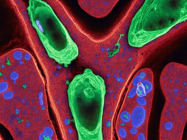
GFP (Green Fluorescent Protein): Enables the identification of tumor cell boundaries in tissue sections and facilitates the separation of tumor cells from infiltrating immune cells during flow cytometry.
Experimental Design: Critical Selection of DBA/2 Hosts
Success hinges on the correct selection of the host strain. Since KLN205 originates from the DBA/2 lineage, DBA/2 mice must be used as recipients. Inoculating these cells into common C57BL/6 mice (H-2b) will trigger a potent allogeneic rejection, clearing the tumor cells and causing experimental failure.
Primary Inoculation Pathways:
Tail Vein Injection: Simulates hematogenous metastasis, where cells colonize the lungs to form diffuse nodules.
Orthotopic Lung Inoculation: Simulates the primary lung tumor microenvironment more accurately.
Subcutaneous Inoculation: Useful for rapid drug screening and accessibility.
Technical Note: Prior to inoculation, it is recommended to remove selection antibiotics (e.g., Puromycin) from the culture medium to prevent non-specific immune stimulation in the host mice.
Efficacy Evaluation: Combining Bioluminescence and Fluorescence
The occult nature of lung tumors makes traditional caliper measurement impossible. KLN205-Luc-GFP solves this through multi-modal quantification:
In Vivo (Luciferase): Following administration of immunotherapeutic agents (e.g., anti-PD-1), longitudinal monitoring is performed using an IVIS system. Responders show a plateau or downward trend in Total Flux (p/s), while non-responders show exponential growth.
Ex Vivo (GFP): At the experimental endpoint, GFP allows for the detection of micrometastases that are invisible to the naked eye. This provides a more sensitive assessment of drug efficacy in preventing metastasis.
Microenvironment Analysis: Correlating Imaging with Flow Cytometry
Tumor burden data alone is insufficient to reveal the mechanisms of immunotherapy. KLN205-Luc-GFP provides a complete "efficacy-mechanism" chain of evidence:
Precise FACS Gating: In Flow Cytometry, the GFP signal acts as a definitive marker to separate tumor cells (GFP+) from host immune cells (GFP-). This eliminates the need for potentially inconsistent tumor-specific surface markers.
TIL Analysis: By gating on the GFP- population, researchers can precisely analyze Tumor-Infiltrating Lymphocytes (TILs), such as the CD8+ T cell to FoxP3+ Treg ratio.
Spatial Distribution: Immunofluorescence (IF) staining of GFP+ tissues allows for the visualization of how CD8+ T cells infiltrate the tumor core versus remaining trapped in the stroma.
Use the KLN205-Luc-GFP cell line to authentically recapitulate tumor-immune interactions, helping you parse CD8+ T cell infiltration and immune escape mechanisms. Order Now>>
References
[1] Kaneko, T., & LePage, G. A. (1978). Growth characteristics and drug responses of a murine lung carcinoma in vitro and in vivo. Cancer Research, 38(7), 2084-2090.
[2] Cumberland, R., et al. (2022). Characterization of the KLN205 Murine Squamous Cell Lung Carcinoma Syngeneic Model. Journal of Immunology, 208 (1 Supplement), 55.12.
[3] Close, D. M., et al. (2010). In vivo bioluminescent imaging (BLI): Noninvasive visualization and interrogation of biological processes in living animals. Sensors, 11(1), 180-206.
[4] Hoffman, R. M. (2015). The multiple uses of fluorescent proteins to visualize cancer cells in vivo. Nature Reviews Cancer, 5(10), 796-806.
