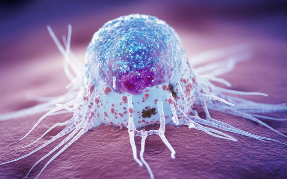
MC38-OVA: An Organoid Co-culture Model in Lung Cancer Study
Research BackgroundLung cancer is one of the most prevalent and lethal malignancies worldwide. Although immune checkpoint inhibitors (ICIs), such as anti-PD-1 (αPD1), have significantly improved outcomes for patients with advanced lung cancer, only a minority derive clinical benefit. Current predictive biomarkers (e.g., PD-L1 expression, tumor mutational burden) lack universal applicability. Furthermore, conventional two-dimensional cell models fail to recapitulate the complex interactions between tumors and the systemic immune system, making it difficult to dynamically track the complete process of T cell infiltration, activation, and killing. While organoid technology has been applied to cancer research, existing co-culture systems inefficiently model the systemic interactions between peripheral immune cells and the tumor microenvironment. Therefore, a novel experimental model that more closely mimics the physiological in vivo state is urgently needed to fill this gap.
Article Information
· Journal: Cell Stem Cell
· Title: An organoid co-culture model for probing systemic anti-tumor immunity in lung cancer
· Affiliations: Tsinghua University, Peking University People's Hospital, Beihang University
· Key Findings: For the first time, a gel-liquid interface (GLI) co-culture model was established using lung cancer organoids (LCOs) and peripheral blood mononuclear cells (PBMCs). This model accurately recapitulates the systemic anti-tumor immune response in lung cancer patients, effectively predicts the efficacy of immune checkpoint inhibitor (ICI) therapy, and reveals the differentiation dynamics of circulating effector memory T (Tem) cells into tumor-reactive T cells. This provides an in vitro platform with both clinical predictive value and potential for mechanistic studies in precision lung cancer immunotherapy.
Experimental Methods
1. Establishment of the GLI Co-culture Model: A superhydrophobic microwell array chip was used. LCOs were mixed with Matrigel and cultured in an inverted configuration, positioning the organoids at the gel-liquid interface. PBMCs were then seeded onto the gel surface to establish the co-culture system. Matrigel concentration (80%) and settling time (10 minutes) were optimized to ensure organoid stability and efficient immune cell infiltration.
2. Model Validation Experiments: Mouse lung cancer (MC38, LLC, MC38-OVA) organoid models were established. Following αPD1 treatment, T cell infiltration and activation markers (IFNγ, CD107a) were assessed using live/dead staining and flow cytometry. The ability of the GLI model to mimic the immune response was compared to in vivo tumor models.
3. Clinical Sample Validation: Tumor tissues and matched PBMCs were collected from 34 treatment-naïve lung cancer patients to establish patient-derived GLI models. Following treatment with αPD1 monotherapy or αPD1 plus chemotherapy, the response index (Ri) was calculated and correlated with patient clinical outcomes (assessed by RECIST criteria).
4. Multi-omics and Functional Analyses: Functional association single-cell RNA sequencing (FascRNA-seq) was used to analyze the dynamic changes in T cell subpopulations. Flow cytometry and quantitative real-time PCR were used to validate the phenotype and function of tumor-reactive T cells. Multiplex immunohistochemistry (mIHC) was used to analyze the distribution of immune cells within the tumor microenvironment.
5. Biomarker Identification: Differential gene expression analysis and clinical correlation were performed to identify characteristic markers of circulating tumor-reactive T cells. The prognostic value of these markers was validated using the TCGA database.
Contact us to discuss your research needs. Our MC38-OVA cell line has gone though a strict QC procedure — request a quote now to get started.
Order now: MC38-OVA cell line, MC38 cell line, LLC cell line

Key Results
Figure 1: Establishment and Validation of the GLI Co-culture Model
This figure illustrates the construction process and core characteristics of the GLI model. After optimization, organoids were stably positioned at the gel-liquid interface, and PBMCs efficiently infiltrated the organoids (significantly better than conventional co-culture methods). Validation in mouse lung cancer models showed that αPD1 treatment induced cell death in MC38 (ICI-sensitive) organoids in the GLI model, but had no significant effect on LLC (ICI-resistant) organoids. This was consistent with the trends in tumor growth inhibition observed in vivo. Additionally, there was increased infiltration of CD8+ T cells and upregulation of activation markers (IFNγ, CD107a), confirming that the model recapitulates ICI-mediated anti-tumor immune responses.

Figure 2: αPD1 Induces Systemic Expansion of Tumor-Reactive T Cells
Experiments in the MC38-OVA mouse model demonstrated that αPD1 treatment significantly suppressed tumor growth. In the GLI model, the proportion of dead organoids and the response index were significantly higher compared to organoid culture alone. Flow cytometry revealed that following αPD1 treatment, the proportion of OVA-specific CD8+ T cells (antigen-specific T cells) significantly increased in tumor tissues, PBMCs, and the GLI model. These cells also highly expressed activation markers such as IFNγ and CD107a, confirming the model's ability to simulate the systemic expansion and activation of tumor-reactive T cells.

Figure 3: GLI Model Accurately Recapitulates Clinical Response to Immunotherapy in Patients
In patient-derived GLI models from 34 lung cancer patients, the response index (Ri) for αPD1 monotherapy and combination therapy was highly consistent with patient clinical outcomes: Ri was significantly higher in patients achieving partial response (PR) compared to those with stable disease (SD) or progressive disease (PD). The predictive accuracy of the GLI model surpassed that of organoid culture alone and conventional PD-L1/TMB biomarkers. Correlation analysis showed a strong correlation between the model Ri and the rate of tumor regression in patients (R=0.8910), confirming its clinical predictive value.

Figure 4: Circulating Tumor-Reactive T Cells Correlate with Immunotherapy Response
Co-culture experiments of PBMCs with autologous organoids showed that the expression levels of IFNγ and CD107a in CD8+ T cells from PBMCs of immune responders (R) were significantly higher than those from non-responders (NR). Furthermore, activated CD8+ T cells enhanced the tumor-killing effect in the GLI model. Co-culture of PBMCs with allogeneic organoids did not produce this effect, confirming that T cell activation is antigen-specific. Moreover, the activation level of circulating T cells significantly correlated with the GLI model Ri, suggesting that circulating tumor-reactive T cells could serve as a predictive biomarker for treatment efficacy.

Figure 5: Differentiation Dynamics from Effector Memory T cells to Tumor-Reactive T cells
Single-cell sequencing analysis revealed that the proportions of effector memory T (Tem) cells and proliferative T (Tprof) cells in PBMCs were significantly higher in responders compared to non-responders, and these cells highly expressed migration-related genes (e.g., ITGB2, ITGB7). Pseudotime trajectory analysis revealed the T cell differentiation path: naive T (Tn) → effector memory T (Tem) → tumor-reactive T (Tt-react) → exhausted T (Tex). Tt-react cells highly expressed cytotoxicity genes (e.g., NKG7, GZMB), and their frequency strongly correlated with the GLI model Ri, confirming that Tem cells are the primary source of tumor-reactive T cells.

Figure 6: Identification of Circulating Tumor-Reactive T Cell Markers
Through differential gene screening and clinical validation, CD9, CD44, and GNLY were identified as core markers of circulating tumor-reactive T cells. Their expression levels in PBMCs and GLI model-infiltrating T cells were significantly higher in responders compared to non-responders. Their expression gradually increased during the differentiation from Tem to Tt-react cells. Analysis of the TCGA database showed that lung cancer patients with high expression of CD9, CD44, and GNLY had significantly longer overall survival, confirming that this marker combination has value for both efficacy prediction and prognosis assessment.

Figure 7: Divergent Molecular Mechanisms Underlying Different Immune Response Patterns
Responders could be categorized into dual responders (DR, response in both organoid and GLI models) and single responders (SR, response only in the GLI model). The DR group exhibited a high proportion of Tprof cells in PBMCs, high expression of stem cell-like and proliferation-related genes (e.g., TCF7, MKI67), and enrichment of myeloid cells (macrophages, dendritic cells) in the tumor microenvironment, recruiting T cells via the CXCL9/10-CXCR3 axis. The SR group predominantly featured Tem cells, highly expressed cytotoxicity genes but were prone to exhaustion, and recruited T cells primarily via the T-T cell CCL5-CCR5 axis. These findings reveal divergent molecular mechanisms underlying different immune response patterns.

This study successfully established a GLI co-culture model of lung cancer organoids and autologous PBMCs. By optimizing the interface design, this model achieves efficient interaction between immune cells and tumor organoids, accurately recapitulating the patient's systemic anti-tumor immune response. Its accuracy in predicting ICI efficacy is significantly superior to traditional biomarkers and organoid culture alone.
The advantages of this model lie in its balance of clinical utility and value for mechanistic studies. It is operationally simple, requires minimal sample, and enables integrated analysis of drug screening, immune dynamic tracking, and biomarker validation.
