Comprehensive Culture Protocol for Human Primary NK Cells: Isolation, Activation, Expansion, Cryopreservation and Functio
Natural Killer (NK) cells are potent innate immune effectors that eliminate tumor cells and virus-infected targets without prior antigen sensitization. Successfully isolating, activating, expanding, and cryopreserving NK cells in vitro is the cornerstone of both basic immunology research and translational cell therapy — yet each step harbors hidden pitfalls that can derail experiments. This comprehensive protocol guide breaks down the entire NK cell culture workflow, from PBMC separation via Ficoll density gradient to long-term cryostorage and revival, so your experiments can proceed exactly as planned.
I. NK Cell Overview
Natural Killer (NK) cells are a subset of lymphocytes belonging to the innate immune system. Unlike T and B cells of the adaptive immune system, NK cells can recognize and destroy pathogen-infected cells and malignant tumor cells without requiring prior antigen-specific priming or clonal expansion. This unique capability makes them front-line defenders in tumor immune surveillance, antiviral immunity, and allogeneic cell therapy applications.
Classical Killing Mechanisms
NK cells eliminate target cells through two well-characterized cytotoxic pathways:
· Perforin–Granzyme Pathway: Upon activation, NK cells release cytoplasmic granules containing perforin (a pore-forming protein) and granzymes (serine proteases). Perforin oligomerizes to form transmembrane pores in the target plasma membrane; granzymes enter through these channels to activate caspase-dependent and -independent apoptosis cascades, leading to rapid target cell death.
· Fas–FasL Death Receptor Pathway: The Fas ligand (FasL, CD95L) expressed on activated NK cells binds to the Fas receptor (CD95) on susceptible target cells. This engagement triggers the formation of the death-inducing signaling complex (DISC), activating caspase-8 and initiating the extrinsic apoptotic pathway.
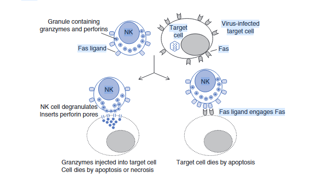
Fig. 1 Schematic illustration of the two classical NK cell cytotoxic killing mechanisms: perforin–granzyme granule exocytosis (left) and Fas–FasL death receptor signaling (right).
Phenotype and Subpopulation Classification
The canonical surface phenotype defining human NK cells is CD3⁻ CD56⁺. The CD3 marker serves as a critical discriminator from NKT cells, which co-express CD3 and CD56 (CD3⁺ CD56⁺). Based on differential expression levels of CD56, human NK cells segregate into two functionally distinct subpopulations:
· CD56ᵇʳᴵᴳᴴₜ (CD56-bright): High CD56 expression with low or absent CD16 (FcγRIIIa). These cells predominantly produce cytokines such as IFN-γ and TNF-α, exerting immunoregulatory functions. They exhibit robust proliferative capacity but comparatively weak direct cytotoxicity, playing key roles in early antiviral responses and maintenance of immune homeostasis.
· CD56ᵈᴵᴹ (CD56-dim): Low CD56 expression with high CD16 expression. This subset possesses potent natural cytotoxic activity and mediates antibody-dependent cellular cytotoxicity (ADCC) through FcγR-mediated recognition of antibody-opsonized targets.
II. NK Cell Isolation and Culture
The isolation and expansion protocol for cord-blood-derived NK cells follows the same principles as peripheral blood sourcing but requires extended culture duration due to the lower initial frequency of CD56⁺ cells in cord blood. The following detailed procedure describes peripheral blood as the starting material.
Step 1: PBMC Isolation by Ficoll Density Gradient Centrifugation
1. Centrifuge fresh anticoagulated whole blood at 2,300 rpm for 10 min at room temperature. Aspirate the upper plasma layer and dilute the remaining blood 1:1 with phosphate-buffered saline (PBS).
2. Carefully layer the diluted blood onto an equal volume of Ficoll-Paque density gradient medium in a conical centrifuge tube. Centrifuge at 2,000 rpm for 20 min with brake off (acceleration 1, deceleration 0).
3. After centrifugation, four distinct layers become visible: the uppermost plasma layer, a translucent buffy coat containing mononuclear cells at the plasma-Ficoll interface, the clear Ficoll separation medium, and a bottom pellet of erythrocytes and granulocytes.
4. Using a sterile pipette, carefully aspirate the buffy-coat PBMC layer and transfer it to a new centrifuge tube. Wash cells twice with PBS at 1,200 rpm for 8 min per wash (acceleration 1, deceleration 0).
5. Resuspend the washed cell pellet in complete medium and perform an accurate total cell count using a hemocytometer or automated cell counter.
Step 2: NK Cell Activation Culture
6. Prepare culture dishes by coating with anti-CD16 soluble antibody solution (or anti-NKp46/anti-CD2 alternatives). Incubate coated dishes overnight at 4°C to allow stable antibody binding.
7. Wash coated dishes 2–3 times with sterile PBS to remove unbound antibody. Add serum-free NK cell expansion medium supplemented with key cytokines including recombinant human IL-2 (typically 100–200 IU/mL) and IL-15 (5–10 ng/mL).
8. Seed freshly isolated PBMCs at a density of 2–2.5 × 10⁶ viable cells/mL into the prepared dishes. Place cultures in a humidified incubator maintained at 37°C with 5% CO₂.
Step 3: NK Cell Expansion Culture
During the first week of expansion, supplementation with 5% heat-inactivated autologous plasma (or AB-type serum substitute) helps maintain optimal cell health, improving both viability and proliferation kinetics. Throughout the expansion period, replenish growth factors and serum-free medium every 48 hours. Monitor cell concentration regularly and adjust the density back to approximately 1 × 10⁶/mL to maintain logarithmic growth conditions.
Step 4: NK Cell Phenotypic and Functional Validation
NK cell identity and purity are confirmed primarily by multicolor flow cytometry using antibodies against CD3 and CD56; a well-expanded preparation should exceed 95% CD3⁻ CD56⁺ purity. Functional potency is assessed using standard chromium-release or flow-cytometric cytotoxicity assays against K562 leukemia target cells across a range of effector-to-target (E:T) ratios.
Skip tedious culture steps, avoid experimental pitfalls, and accelerate your immunology and cell therapy projects with our reliable NK cells—start your research with premium NK cells now!
Order Now: NK-92 Cell Line |NK-92MI Cell Line
III. NK Cell Cryopreservation and Thawing
Cryopreservation Procedure
A cryoprotectant formulation containing 10% DMSO (dimethyl sulfoxide) is recommended to protect cell membranes from ice crystal damage during freezing. Collect expanded NK cells by gentle centrifugation (300 ×g, 5 min), discard supernatant, and resuspend the pellet in pre-chilled cryopreservation medium at a density of 1 × 10⁶ to 1 × 10⁷ cells/mL. Transfer aliquots into cryovials and subject them to controlled-rate cooling (approximately −1°C/min) before transferring vials to a −80°C freezer overnight, followed by long-term storage in liquid nitrogen vapor phase. For serum-free workflows, dedicated serum-free immune-cell cryopreservation media are commercially available as validated alternatives.
Thawing / Revival Procedure
Pre-warm a water bath to 37°C and have complete culture medium ready and warmed to the same temperature. Retrieve the cryovial from liquid nitrogen storage, place it inside a sealed PE glove (for biosafety), and immerse the glove in the 37°C water bath while gently agitating until all visible ice has melted. Immediately transfer the thawed cell suspension into a 15 mL conical tube containing 5 mL of warm medium. Centrifuge at 300 ×g for 5 min to remove DMSO-containing cryoprotectant, discard supernatant, and gently resuspend the cell pellet in fresh complete medium. Transfer to a pre-warmed culture vessel and place it undisturbed in a 37°C, 5% CO₂, humidified incubator.
IV. Representative Experimental Data
Human primary NK cells expanded using an optimized antibody–cytokone co-stimulation protocol display characteristic clustered morphology under phase-contrast bright-field microscopy, reflecting their activated state and high viability (Fig. 2). Multicolor flow cytometric analysis confirms that the expanded population achieves >95% purity with a canonical CD3⁻ CD56⁺ phenotype , demonstrating effective enrichment over the starting PBMC population. In functional validation assays, these expanded NK cells exhibit potent dose-dependent cytotoxic activity against K562 chronic myeloid leukemia target cells across effector-to-target ratios spanning 50:1 to 5:1, confirming robust post-expansion effector functionality suitable for downstream research or therapeutic development applications.
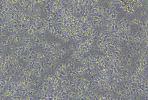
Fig.2 Bright-field micrograph (×100 magnification) of human NK cells after 14 days of antibody–cytokine activation and expansion, demonstrating typical cluster morphology indicative of healthy, activated NK cell growth.
Explore Vitrobiotech's complete portfolio of validated NK cell products, serum-free expansion systems
Order Now: NK-92 Cell Line |NK-92MI Cell Line
