
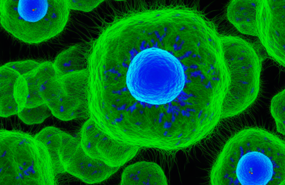
OVA Cells| How neutrophil and CAR macrophage fusion syncytial cells enhance solid tumor immunotherapy
In 2026, Tianyi Tian, Siyu Zhao, Tian Tian, et al. published "Neutrophil integrated syncytial CAR macrophage for cancer immunotherapy" in Nature Immunology. This study fuses second-generation CAR macrophages with neutrophils to construct syncytial CAR macrophages (S-CAR-M). The fused cells inherit CXCR2-mediated tumor chemotaxis and NET/ROS effector functions while retaining CAR recognition, phagocytosis, and antigen presentation capabilities. In multiple mouse solid tumor models, S-CAR-M expands intratumoral residence, clears tumors via dual scFv-antigen and PtdSer-MerTK pathways, and induces host T cell antigen spreading.
01 Problem Identification: Chemotherapy, radiotherapy, surgery, and SMAD4 deficiency all increase tumor neutrophil recruitment. Based on this, the study transfers the migratory capacity of neutrophils to CAR-M.
02 Fusion Phenotype: S-CAR-M simultaneously expresses Ly6G, F4/80, and CAR, retaining CXCR2, NET formation programs, and macrophage immune functions.
03 Killing Mechanism: DNase I, NAC, and MerTK inhibition experiments link NET, ROS, PtdSer exposure, and tumor debris phagocytosis.
04 In Vivo Efficacy: Liver metastasis, peritoneal metastasis, post-radiotherapy, post-surgical recurrence, and human xenograft models collectively test tumor suppression and survival.
05 Immune Spreading: Bilateral heteroantigen tumors, OVA tetramers, rechallenge, and single-cell sequencing support host CD8 T cell antigen spreading.
Based on the article, here is a summary of the cell lines used, what each was used to validate, and the phenomena they demonstrated.
I. Murine Cell Lines
1. MC38 (Mouse Colon Cancer Cells)
· Validation: Used as the core syngeneic tumor model to generate MC38-hHER2 (exogenous human HER2 expression), MC38-OVA (OVA antigen expression), and MC38-hHER2/OVA derivatives. Employed in liver metastasis models (including SMAD4-deficient variants), subcutaneous tumor models, bilateral heteroantigen tumor models, antigen-mixed models, rechallenge experiments, and single-cell RNA sequencing.· Phenomena Demonstrated: S-CAR-M showed higher entry into liver metastases at 24 hours than CAR-M and maintained this up to 72 hours; it completely cleared liver metastases in 5/8 mice and improved survival. In the bilateral heteroantigen model, S-CAR-M simultaneously suppressed the target-positive tumor and the contralateral hHER2-negative MC38, an effect that disappeared in immunodeficient mice, indicating that antigen spreading depends on the host immune system. MC38 conditioned medium was also used for in vitro chemotaxis assays.
2. CT26 (Mouse Colon Cancer Cells)
· Validation: After oxaliplatin chemotherapy, used to detect changes in intratumoral Ly6G-positive neutrophils and CXCR2 expression; also used as a tumor model for S-CAR-M infusion after chemotherapy.· Phenomena Demonstrated: Oxaliplatin treatment increased intratumoral neutrophils and CXCR2 signaling, demonstrating that standard chemotherapy can create a chemotactic window; S-CAR-M showed higher tumor entry proportions than CAR-M in the post-chemotherapy CT26 model.
VITRO BIOTECH also offers CT26-OVA (OVA antigen expression) model for the immunotherapy studies.
3. 4T1 (Mouse Breast Cancer Cells)
· Validation: 4T1-hHER2 was generated for post-radiotherapy models and incomplete resection/post-surgical residual models to evaluate tumor control and survival benefit of S-CAR-M.· Phenomena Demonstrated: Both radiotherapy and incomplete resection increased intratumoral neutrophil recruitment; CXCL1 peaked on day 9 after low-dose radiotherapy, and S-CAR-M infusion at this time point delayed 4T1-hHER2 tumor growth and prolonged survival. In the post-surgical residual model, S-CAR-M reduced local recurrence (3/7 mice achieved sustained remission) and reduced lung metastatic nodules.
4T1-OVA cell line is available in stock. Contact us now for more details>>
4. B16-F10-OVA (Mouse Melanoma Cells, Mentioned in Figure Section)
· Validation: Used in the relevant figure to validate the anti-tumor efficacy of hydrogel encapsulating OVA-specific CD8+ T cells and IL-15 (Note: this belongs to a separate hydrogel study cited in the article, presented alongside the main S-CAR-M study).· Phenomena Demonstrated: Peritumoral injection of hydrogel encapsulating OVA-specific CD8+ T cells and IL-15 significantly inhibited tumor growth, with >90% tumor volume reduction in the high-dose group.
5. RAW264.7 (Mouse Macrophage Cell Line)
· Validation: Used as the cellular background in the RIMTAC study (a separate JMC paper cited in the article) to validate compound 10-induced BRD4 degradation (this belongs to the cited RIMTAC platform study, not the main S-CAR-M study). Not used as a primary validation cell in the S-CAR-M study.6. LLC (Mouse Lewis Lung Carcinoma Cells)
· Validation: Mentioned in the context of the LLC-Luc allograft lung metastasis model (this belongs to the BJ-2412 study section). Not used as a core model in the main S-CAR-M study.
LLC-OVA model is also available.
II. Human Cell Lines
1. HT-29-hHER2 (Human Colon Cancer Cells, Exogenous hHER2 Expression)
· Validation: Used to establish xenograft models for human hS-CAR-M (CAR-THP-1 macrophages fused with peripheral blood neutrophils).· Phenomena Demonstrated: hS-CAR-M outperformed hCAR-M, with 1/6 mice achieving complete remission, demonstrating the feasibility of human fused cells, though still at the xenograft proof-of-concept stage.
2. BT-474 (Human Breast Cancer Cells)
· Validation: Used in extended experiments to validate killing by hS-CAR-M constructed from primary human macrophages.· Phenomena Demonstrated: Further supported the killing capacity of human fused cells, but did not provide cross-donor or cross-batch consistency data.
3. THP-1 (Human Acute Monocytic Leukemia Cell Line)
· Validation: Differentiated into macrophages to construct CAR-THP-1 macrophages, which were then fused with peripheral blood neutrophils to form human hS-CAR-M; also used in tumor-macrophage co-culture systems (this part is related to the cited BJ-2412 study).· Phenomena Demonstrated: THP-1-derived hS-CAR-M showed efficacy in the HT-29-hHER2 xenograft model, but THP-1-derived and primary macrophage-derived products have different characteristics that may affect fusion, migration, and NET release.
III. Other Cell Types
1. Mouse Bone Marrow-Derived Macrophages (BMDM)
· Validation: Used to construct second-generation anti-hHER2 CAR-M, which were then PEG-fused with neutrophils and flow-sorted to obtain S-CAR-M. These are the core starting cells of the main S-CAR-M study.· Phenomena Demonstrated: After fusion, S-CAR-M simultaneously expressed Ly6G, F4/80, and CAR, retaining CXCR2, NET formation programs, and macrophage immune functions (CD80, CD86, MHC II, CD40, MerTK, CD64, etc.).
2. Neutrophils (Murine and Human Peripheral Blood Sources)
· Validation: Fused with CAR-M or CAR-THP-1 macrophages; also used as controls for NET release experiments.· Phenomena Demonstrated: Neutrophils enter post-treatment inflammatory foci along the CXCL-CXCR2 axis and release NETs and ROS; after fusion, S-CAR-M released MPO- and H3Cit-positive NET structures with DNA release approaching that of neutrophils, but retained macrophage nuclei and basic morphology, distinct from typical suicidal NETosis.
Overall, the study systematically validated the chemotaxis, residence, killing, antigen spreading, and safety of S-CAR-M using murine syngeneic models (MC38, CT26, 4T1), preliminarily validated clinical translation feasibility using human cells (HT-29-hHER2, BT-474, THP-1), while neutrophils and BMDM served as the core cell sources for constructing S-CAR-M.
Reference
Neutrophil-integrated syncytial CAR macrophage for cancer immunotherapy. DOI:10.1038/s41590-026-02615-2
