Decoding the Raji Cell Line: Why It Became the Gold Standard in Immunology Research
Introduction
In rigorous scientific inquiry, a widely validated and accepted cell line functions like a chemical standard in metrology; it provides a fundamental basis for ensuring the comparability and reproducibility of results across different laboratories and studies. Within the fields of immunology and B-cell malignancy research, the Raji cell line, derived from a human Burkitt's lymphoma, perfectly embodies this role. Since its establishment in 1963, its unique biological characteristics have made it an indispensable tool for countless laboratories worldwide in the development and evaluation of immunotherapies.
Eliminate uncertainty in your experimental outcomes. We provide rigorously STR-profiled and quality-controlled Raji cells to ensure the reliability of your data. Order Now>>
A Stable B-Cell Phenotype
One of the most significant attributes of the Raji cell line is its consistent, high-level expression of key B-lymphocyte surface antigens. These molecular markers, such as CD19, CD20, and CD22, act as definitive identifiers for B-cells. Critically, these proteins are the primary targets for numerous modern immunotherapies, particularly monoclonal antibodies and Chimeric Antigen Receptor (CAR) T-cell therapies.
For instance, anti-CD20 monoclonal antibodies like rituximab are a cornerstone of B-cell lymphoma treatment. During the early stages of drug development, researchers require a reliable in vitro model to confirm that a newly developed antibody can accurately recognize and bind to its CD20 target. Raji cells, with their high CD20 expression, serve as the ideal positive control for this purpose. Techniques such as flow cytometry can be used to easily quantify the binding efficiency of the antibody to Raji cells. Similarly, when developing CAR-T cells targeting CD19, Raji cells are used as target cells to test whether the CAR-T cells' recognition and binding capabilities meet design specifications. This stable phenotype provides a uniform and dependable standard for the initial screening and functional validation of immunotherapies.
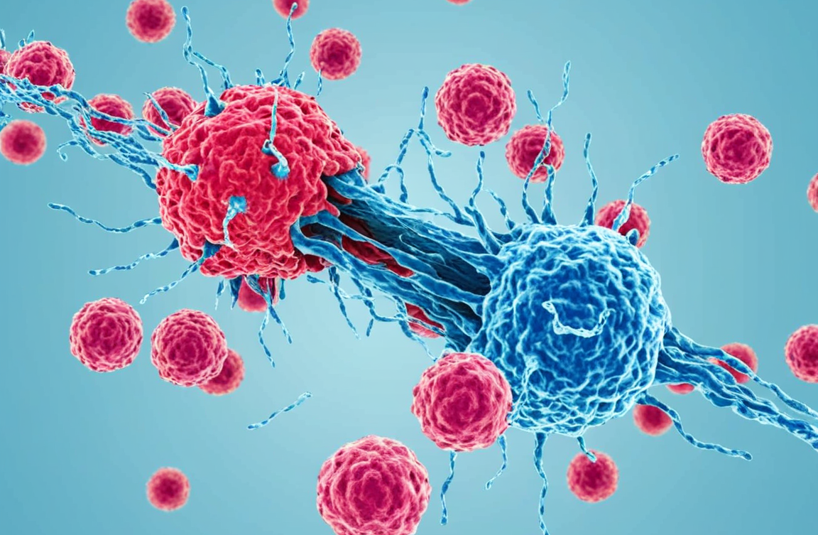
Sensitivity and Reproducibility in Immune-Mediated Killing
Beyond serving as a recognition target, Raji cells exhibit a clear and highly reproducible response to various immune-mediated killing mechanisms. This quality makes them the ideal model for establishing and standardizing cytotoxicity assays. This is primarily demonstrated in two key assays:
Antibody-Dependent Cell-Mediated Cytotoxicity (ADCC): When an anti-CD20 antibody (e.g., rituximab) binds to the CD20 antigen on the surface of a Raji cell, the antibody's Fc region engages with the FcγRIIIa (CD16a) receptor on Natural Killer (NK) cells. This engagement activates the NK cell, prompting it to release cytotoxic granules like perforin and granzymes, which ultimately induce lysis of the Raji cell. Because Raji cells respond consistently in this process, they are widely used as the "standard target cell" for evaluating the potency of novel anti-CD20 antibodies in inducing ADCC.
Complement-Dependent Cytotoxicity (CDC): Certain antibodies (like IgM or specific IgG subclasses), upon binding to antigens on the Raji cell surface, can activate the complement cascade in serum. This cascade culminates in the formation of a Membrane Attack Complex (MAC) on the cell membrane, leading to pore formation and osmotic lysis. Raji cells are also sensitive to CDC, making them a common tool for measuring the ability of an antibody therapeutic to activate the complement system.
This predictable responsiveness allows researchers to accurately compare the cytotoxic efficacy of different drugs or immune effector cells, ensuring the reliability of experimental outcomes.
A Defined Genetic Background and Extensive Historical Data
As one of the first human lymphocyte cell lines ever established, Raji has been a subject of scientific study for over half a century. This long history has resulted in a massive repository of published literature and data. Its genomic, transcriptomic, and proteomic information is well-documented and readily available in public databases.
Its defined genetic background, which includes the characteristic c-MYC gene translocation t(8;14)(q24;q32) that is central to the pathogenesis of Burkitt's lymphoma, makes it a classic model for studying the molecular biology of this malignancy. This greatly facilitates experimental design, interpretation of results, and comparison with other studies, mitigating the uncertainty that can arise from using cell lines with poorly characterized backgrounds.
Accelerate your immuno-oncology research with a stable B-cell phenotype and a dependable immune response. Take the first step to success with our validated Raji cells. Learn more>>
Conclusion
In summary, the status of the Raji cell line as a "gold standard" in immunology research is no coincidence. Its stable expression of key B-cell antigens makes it an ideal positive control for therapeutic development; its clear and reproducible response to immune-killing mechanisms like ADCC and CDC makes it a reliable target for functional assays; and its extensive research history and defined genetic background provide robust data support for scientific investigation. It is the combination of these three core features—a stable phenotype, a reliable response, and a wealth of data—that has cemented Raji's indispensable position as a benchmark tool in immunology and oncology research.
References
[1]Dall'Ozzo, S., et al. (2004). A new, simple and rapid non-radioactive assay for antibody-dependent-cell-mediated cytotoxicity. Journal of Immunological Methods, 291(1-2), 191-203.
[2]Bindon, C. I., et al. (1988). Importance of antigen density and antibody class in the complement-dependent lysis of target cells. Immunology, 65(3), 437–444.
[3]Pulvertaft, R. J. V. (1964). Cytology of Burkitt's Tumour (African Lymphoma). The Lancet, 283(7327), 238-240.
[4]Dalla-Favera, R., et al. (1982). Human c-myc onc gene is located on the region of chromosome 8 that is translocated in Burkitt lymphoma cells. Proceedings of the National Academy of Sciences, 79(24), 7824-7827.
