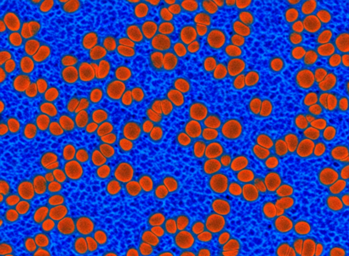
SW839 Cells: A Fundamental Model for Exploring Colorectal Cancer Cell Proliferation, Apoptosis, and Invasion
Introduction
Rectal cancer is one of the most common malignant tumors worldwide, characterized by a complex pathogenesis involving multi-gene and multi-step evolution. A profound understanding of the fundamental biological behaviors of rectal cancer cells, such as uncontrolled proliferation, resistance to apoptotic signals, and invasive/metastatic capabilities, is crucial for developing novel diagnostic markers and therapeutic strategies. In vitro cultured cancer cell lines, owing to their ease of manipulation, good reproducibility, and relatively low cost, have become indispensable tools in cancer biology research. The SW839 cell line, derived from a primary rectal adenocarcinoma of a 72-year-old female (Fogh & Trempe, 1975), provides researchers with a valuable platform to simulate and investigate key characteristics of rectal cancer cells at molecular and cellular levels, thereby laying the groundwork for more complex in vivo studies and clinical translation.
Application of SW839 Cell Line in Studying Cell Proliferation and Cell Cycle
Uncontrolled cell proliferation is one of the most fundamental characteristics of cancer. The SW839 cell line is frequently used to investigate intrinsic and extrinsic factors affecting rectal cancer cell proliferation and the regulatory mechanisms of the cell cycle.
1. Growth Curve Determination and Proliferation Assessment
Researchers can determine the growth curve of SW839 cells using various methods to assess their basal proliferation rate or the impact of different treatments (e.g., drugs, gene overexpression/knockdown) on proliferation. Common methods include:
MTT/CCK-8 Assay: Based on the activity of mitochondrial dehydrogenases in viable cells, this colorimetric assay quantifies the number of living cells. It is a simple, high-throughput method widely used for drug screening and cell viability testing (Mosmann, 1983).
Direct Cell Counting: Using a hemocytometer or an automated cell counter to directly count cell numbers.
BrdU/EdU Incorporation Assay: By detecting the incorporation of thymidine analogs (BrdU or EdU) during DNA replication, this method accurately assesses the proportion of S-phase cells and proliferative activity.
2. Cell Cycle Analysis
Cyclins, Cyclin-Dependent Kinases (CDKs), and their inhibitors (CKIs) are core regulators of the cell cycle. Flow cytometry analysis of PI (propidium iodide)-stained SW839 cells' DNA content can determine the distribution of cells in G0/G1, S, and G2/M phases. This helps investigate how specific genes or compounds affect the cell cycle progression of SW839 cells, such as inducing G1 arrest or M-phase arrest (Darzynkiewicz et al., 2004).
Need a reliable rectal adenocarcinoma model? Choose our high-quality SW839 cells for a stable in vitro platform in your cancer research. Click to order>>
Investigating Apoptosis and Survival Signals Using the SW839 Cell Line
Apoptosis (programmed cell death) is an essential mechanism for eliminating damaged or superfluous cells, and tumor cells often evade apoptosis through various pathways. The SW839 cell line provides a convenient model for studying the regulation of apoptosis in rectal cancer cells.
Induction and Detection of Apoptosis:
Researchers can induce apoptosis in SW839 cells through various means, such as chemotherapeutic drugs (e.g., 5-FU, oxaliplatin), death receptor agonists (e.g., TRAIL), or overexpression of specific genes.
Methods for detecting apoptosis are diverse, including:
1. Annexin V/PI Double Staining: Annexin V binds to phosphatidylserine exposed on the outer leaflet of the plasma membrane during early apoptosis, while PI can enter late apoptotic or necrotic cells and stain their DNA. Flow cytometry or fluorescence microscopy can distinguish between normal, early apoptotic, late apoptotic, and necrotic cells.
2. Caspase Activity Assay: Caspase family proteins are key executioner enzymes in apoptosis. The occurrence of apoptosis can be assessed by measuring the activity of Caspase-3, -8, -9, etc.
3. TUNEL Assay: Detects the 3'-OH ends generated by DNA fragmentation during apoptosis.
Preliminary Observation of Cell Migration and Invasion Capabilities Using SW839 Cells
Tumor invasion and metastasis are the main causes of cancer-related deaths. The SW839 cell line is also commonly used for preliminary assessment of the motility and basement membrane penetration ability of rectal cancer cells.
Wound Healing Assay:
A "scratch" is artificially created in a confluent monolayer of SW839 cells. The ability of cells to migrate into the scratch area and close the "wound" is observed microscopically over time. This is a simple and intuitive method for assessing 2D cell migration (Liang et al., 2007).
Transwell Invasion Assay:
The bottom of the upper chamber of a Transwell insert is coated with a layer of Matrigel, which mimics the extracellular matrix. SW839 cells are seeded in the upper chamber, and culture medium containing chemoattractants is added to the lower chamber. After a period of incubation, the number of cells that have invaded through the Matrigel and migrated to the lower surface of the membrane is counted to assess the invasive potential. If Matrigel is not used, the assay measures migration ability.
Value of SW839 Cell Line in Basic Chemotherapy Drug Sensitivity Testing
Assessing the sensitivity of cancer cells to chemotherapeutic drugs is an important part of preclinical research. The SW839 cell line is often used as a testing platform.
Determination of IC50 Value:
SW839 cells are exposed to a range of concentrations of chemotherapeutic drugs (e.g., fluorouracil (5-FU), oxaliplatin, irinotecan, commonly used for colorectal cancer treatment). After a certain incubation period, cell viability is assessed using MTT or CCK-8 assays. By plotting a drug concentration-cell viability curve, the half-maximal inhibitory concentration (IC50 value) can be calculated. This value reflects the sensitivity of the cells to the drug; a lower IC50 value indicates higher sensitivity.
Combination Drug Studies:
SW839 cells can also be used for preliminary exploration of the synergistic, antagonistic, or additive effects of combining different chemotherapeutic drugs or combining chemotherapy with other therapeutic modalities (e.g., targeted drugs, radiotherapy).
Investigating cell migration and invasion mechanisms? The SW839 cell line supports wound healing and Transwell assays, vividly demonstrating cellular behavior. Learn more>>
Conclusion: SW839 Provides a Fundamental Platform for Understanding Rectal Cancer Biology
The SW839 cell line, as a classic cell model derived from human rectal adenocarcinoma, offers researchers a convenient and valuable tool for exploring core biological behaviors of rectal cancer cells, such as proliferation, apoptosis, and migration/invasion, as well as for performing preliminary drug sensitivity assessments. Despite certain limitations, through rigorous experimental design, cautious interpretation of results, and validation with other cell models, animal models, and even clinical samples, research findings from the SW839 cell line can provide important fundamental information and clues for a deeper understanding of rectal cancer pathogenesis and the development of novel therapeutic strategies.
References
[1] Fogh, J., & Trempe, G. (1975). New human tumor cell lines. In J. Fogh (Ed.), Human tumor cells in vitro (pp. 115-159). Plenum Press.
[2] Mosmann, T. (1983). Rapid colorimetric assay for cellular growth and survival: application to proliferation and cytotoxicity assays. Journal of Immunological Methods, 65(1-2), 55–63.
[3] Darzynkiewicz, Z., Huang, X., & Okafuji, M. (2004). Cytometric methods to detect apoptosis. Methods in Molecular Biology, 282, 69-87.
[4] Liang, C. C., Park, A. Y., & Guan, J. L. (2007). In vitro scratch assay: a convenient and inexpensive method for analysis of cell migration in vitro. Nature Protocols, 2(2), 329–333.
[5] Valeriote, F., & Lin, H. S. (1975). Synergistic interaction of anticancer agents: a cellular perspective. Cancer Chemotherapy Reports. Part 1, 59(5), 895–900.
[6] Freshney, R. I. (2010). Culture of animal cells: A manual of basic technique and specialized applications (6th ed.). Wiley-Blackwell.
