Tracking Bladder Cancer Metastasis via T24-Luc Bioluminescence
Introduction:
The T24 cell line is a common model derived from human bladder carcinoma. It exhibits high grade and invasive behavior. Researching metastasis requires tools to monitor tumor cells as they migrate from primary sites to distant organs. Traditional methods, such as manual palpation or anatomical inspection, fail to detect early spread. The T24-Luc cell line, engineered with the firefly luciferase gene, addresses this challenge. This modification allows researchers to visualize tumor cells in living subjects without invasive surgery. By emitting light through enzymatic reactions, these cells provide a signal that reveals their exact location and mass. This introduction explores how T24-Luc technology facilitates the study of metastatic progression in bladder cancer research.
Our T24 cell line provides stable growth and high invasiveness for bladder cancer research. It helps you construct high-grade tumor models with consistent experimental results. View Details>>
Understanding Metastatic Potential in T24 Cells
The T24 cell line is known for its motility. In mouse models, these cells spread to the lungs, liver, and lymph nodes. This invasive behavior mirrors the clinical progression of late-stage bladder cancer. To study this spread, researchers need a reliable method to confirm where cells settle after leaving the bladder. T24-Luc cells provide a solution by acting as internal beacons. When these cells enter the bloodstream, they eventually colonize distant tissues. The luciferase marker remains stable during these transitions, ensuring that the daughter cells in metastatic nodules continue to emit signals.
Detecting Small Metastatic Nodules
Bioluminescence imaging (BLI) offers high sensitivity. The luciferase enzyme catalyzes the substrate, D-luciferin, to produce photons. These photons penetrate through animal tissues and are captured by sensitive cameras. This process detects small clusters of cells that are invisible to the naked eye. In early metastasis, a few thousand cells may seed in the lung. Standard imaging like X-ray or ultrasound often misses these clusters due to low resolution. T24-Luc imaging identifies these signals early, allowing for the study of the "pre-metastatic" niche. This detection limit is crucial for evaluating drugs that aim to prevent tumor seeding rather than just shrinking large masses.
Spatiotemporal Tracking of Tumor Spread
One primary advantage of T24-Luc cells is the ability to conduct longitudinal studies. Researchers can monitor the same animal over weeks. This spatiotemporal tracking shows the journey of the cancer. For example, after a tail-vein injection, the initial signal may concentrate in the lungs. Over time, the signal may appear in the bone or brain. Because the animal remains alive, researchers collect data at multiple time points. This reduces the number of animals needed for an experiment. It also eliminates variations between different groups of mice. The resulting growth curves provide a clear picture of how fast the cancer spreads under specific conditions.
Validating Signals with Ex-Vivo Imaging
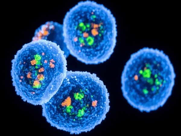
While live imaging is useful, validating the source of the signal is necessary. At the end of the study, organs are removed for ex-vivo imaging. Placing the lungs, liver, and kidneys directly in the imaging chamber confirms the presence of T24-Luc cells. This step removes interference from skin or fur. It allows for the detection of even weaker signals in deep tissues. Following ex-vivo imaging, these tissues are often processed for histology. Because T24-Luc cells are often dual-labeled with fluorescent proteins like GFP, researchers can use fluorescence microscopy to see individual cells within the tissue architecture. This correlation between whole-body signals and microscopic evidence strengthens the experimental conclusions.
Data Analysis and Quantification
Quantifying metastasis requires measuring the "Total Flux" of photons. This value represents the number of photons emitted per second. Unlike diameter measurements, Total Flux reflects the metabolic activity and viable cell count of the tumor. In a metastatic model, an increase in Total Flux over time indicates successful colonization and growth. If a treatment is effective, the signal intensity will plateau or decrease. Researchers must use a consistent ROI (Region of Interest) to compare data across different animals. This standardized approach ensures that the results are reproducible and objective.
Use T24-Luc cells for non-invasive bioluminescence imaging to monitor tumor growth in real-time. This allows you to obtain precise growth curves without sacrificing animals, increasing research efficiency. Learn more>>
Conclusion
The T24-Luc cell line is an essential tool for bladder cancer research. It bridges the gap between primary tumor observation and complex metastasis tracking. By providing real-time, sensitive, and quantifiable data, it allows for a deeper understanding of how bladder cancer spreads. This technology supports the development of new therapies targeting the metastatic process.
References:
[1]Bubeník, J., et al. (1970). Established cell line of urinary bladder carcinoma (T24) containing tumour-specific antigen. International Journal of Cancer, 5(3), 310-319.
