SHP-77 and SHP-77-luc Cell Line: From SCLC Characteristics to In Vivo Imaging Models
Introduction:
Small Cell Lung Cancer (SCLC) is a highly aggressive neuroendocrine malignancy characterized by a short doubling time and early systemic metastasis. In preclinical research, selecting appropriate cellular models is critical for elucidating pathogenesis and screening therapeutics. The SHP-77 cell line, a classic model derived from SCLC patients, possesses distinct "variant" biological characteristics, making it a key tool for studying chemotherapy resistance and tumor heterogeneity. To overcome the limitations of traditional models in monitoring real-time tumor dissemination, the genetically engineered SHP-77-luc luciferase reporter strain was developed. This article will analyze the foundational properties of the parental SHP-77 and explore the advantages of SHP-77-luc in constructing visualizable animal models.
Origin and Background: Representative of Variant SCLC
The SHP-77 cell line was originally isolated from a mediastinal lymph node metastasis in a male patient with SCLC who had not received prior chemotherapy. Unlike classic SCLC cell lines (such as NCI-H69), SHP-77 is classified as "variant" small cell lung cancer (SCLC-v).
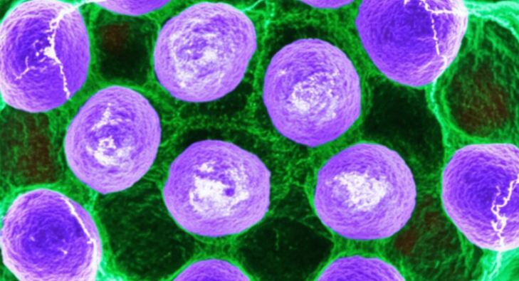
This classification is based on its unique morphological and growth kinetics. Under in vitro culture conditions, SHP-77 cells do not form the tight, spherical floating aggregates typical of classic SCLC; instead, they present as loose suspension aggregates or irregular single-cell suspensions. This morphological difference is often accompanied by a faster cell doubling rate and higher cloning efficiency. Understanding this background is crucial for researchers, as variant SCLC often exhibits primary or secondary resistance to radiation therapy and certain chemotherapeutic agents, rendering SHP-77 an ideal model for studying refractory lung cancer.
Utilize the SHP-77 cell line to construct classic variant SCLC models, precisely elucidating c-myc amplification-driven malignant proliferation mechanisms. Order now>>
Genetic Profile: c-myc Amplification and Neuroendocrine Markers
From a molecular genetics perspective, the most significant feature of the SHP-77 cell line is the amplification and overexpression of the c-myc proto-oncogene. Aberrant activation of c-myc drives rapid cell cycle progression, directly correlating with its highly malignant proliferative phenotype. In contrast, classic SCLC typically presents with L-myc or N-myc amplification.
Furthermore, although SHP-77 retains neuroendocrine properties, the expression levels of certain biochemical markers (such as L-Dopa decarboxylase, L-DDC) are lower compared to classic cell lines. This molecular heterogeneity authentically recapitulates the complexity of tumor tissues in clinical SCLC patients. Utilizing SHP-77 for experiments helps reveal the specific mechanisms by which the c-myc signaling pathway drives tumor metabolic reprogramming and immune escape.
Engineering: Principles of SHP-77-luc Construction
To meet the demand for data quantification in in vivo efficacy evaluations, researchers constructed the SHP-77-luc derivative via lentiviral transduction technology. This process involves stably integrating the gene encoding Firefly Luciferase into the parental genome and selecting high-expressing monoclonal populations using antibiotics (typically Puromycin).
The core value of SHP-77-luc lies in translating biological processes into optical signals. Upon injecting the substrate D-Luciferin into tumor-bearing mice, intracellular luciferase in living tumor cells catalyzes the oxidation of the substrate, releasing photons with a wavelength of approximately 560 nm. This light signal penetrates mammalian tissue and is captured by high-sensitivity imaging systems like IVIS. The intensity of the Photon Flux correlates strictly linearly with the number of viable cells, thereby enabling absolute quantification of tumor burden.
Model Value: From In Vitro Screening to In Vivo Tracing
SHP-77 and SHP-77-luc play complementary roles in translational medicine research, suitable for distinct experimental scenarios.
In Vitro Mechanism Studies (SHP-77):
The parental SHP-77 cell line offers a clean background without interference from exogenous genes, making it the premier choice for High-throughput Screening (HTS) and gene editing (e.g., CRISPR/Cas9 knockout). Researchers utilize its suspension growth characteristics to rapidly assess IC50 values of first-line chemotherapeutics like Etoposide and Cisplatin, or to investigate the molecular mechanisms of c-myc inhibitors.
In Vivo Metastasis Tracing (SHP-77-luc):
SCLC is prone to metastasize to the liver, bones, and brain. Traditional subcutaneous tumor models fail to simulate this process, and visceral tumors cannot be measured via calipers. Using SHP-77-luc to construct tail vein or intracardiac injection models allows researchers to perform longitudinal monitoring of the entire disease course in single mice. Bioluminescence imaging can detect minimal metastatic lesions in the liver or bone marrow early on, even without palpable surface masses. This non-invasive technology not only significantly improves data precision but also reduces the number of animals sacrificed, aligning with the 3R principles of animal ethics.
Rely on the stable bioluminescent signals of SHP-77-luc to realize non-invasive, precise quantification of tumor burden throughout the entire disease course in single mice. Learn more>>
References
[1]Fisher, E. R., & Paulson, J. D. (1985). A new in vitro cell line established from human small cell lung cancer. Cancer Research, 45(11), 5684-5690.
[2]Gazdar, A. F., et al. (1985). Characterization of variant subclasses of cell lines derived from small cell lung cancer having distinct biochemical, morphological, and growth properties. Cancer Research, 45(6), 2924-2930.
[3]Little, C. D., et al. (1983). Amplification and expression of the c-myc oncogene in human lung cancer cell lines. Nature, 306(5939), 194-196.
[4]Meuwissen, R., et al. (2003). Mouse models for small-cell lung cancer. Oncogene, 22(42), 6553-6560.
