NCI-H460 Cell Line : From Large Cell Lung Cancer Characteristics to Multimodal Imaging Models
Introduction:
Non-Small Cell Lung Cancer (NSCLC) accounts for over 85% of all lung cancer cases, with Large Cell Carcinoma (LCC) characterized by poor prognosis due to its undifferentiated nature and aggressive behavior. The NCI-H460 cell line, established in 1982 as a classic human LCC model, has become a core tool for basic lung cancer research and anti-tumor drug screening due to its defined genetic background and rapid growth kinetics. To meet the demands of translational medicine for in vivo visualization and single-cell analysis, researchers developed the NCI-H460-luc cell (bioluminescence-labeled) and NCI-H460-luc-gfp cell (dual-labeled) variants. This article will systematically analyze the biological properties of the parental line and detail how these engineered variants enhance data dimension and precision.
Origin and Molecular Genetic Characteristics
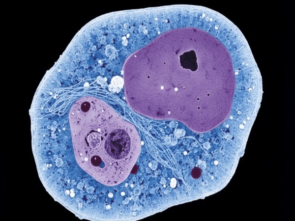
The NCI-H460 cell line was originally isolated from the pleural effusion of a male patient with large cell lung cancer. Pathologically, it presents an undifferentiated epithelial morphology, lacking the typical structural features of adenocarcinoma or squamous cell carcinoma.
From a molecular genetics perspective, NCI-H460 possesses two key genetic features that secure its unique position in pharmacological research:
1. KRAS Mutation: This cell line harbors a heterozygous KRAS Q61H mutation. As a downstream effector of the RAS/MAPK signaling pathway, constitutive activation of KRAS drives tumor proliferation and survival. This makes NCI-H460 an ideal model for evaluating MEK inhibitors, PI3K inhibitors, and drugs targeting KRAS variants other than G12C.
2. p53 Wild-Type: Unlike many lung cancer cell lines (such as NCI-H1299) that harbor p53 deletions or mutations, NCI-H460 retains the wild-type p53 gene. This characteristic renders it relatively sensitive to DNA damage-inducing agents (e.g., Cisplatin, Doxorubicin) and makes it frequently used to study MDM2-p53 interactions and apoptosis mechanisms.
As a classic p53 wild-type model, the NCI-H460 cell line is the premier tool for studying DNA damage repair mechanisms and MDM2-p53 interactions. Order now>>
Growth Kinetics and Tumorigenic Potential
Under in vitro culture conditions, NCI-H460 exhibits an exceptionally high proliferation rate, with a doubling time of approximately 18 to 24 hours. Cells grow adherently, displaying polygonal or spindle shapes, and tend to pile up upon reaching confluence. This rapid division requires researchers to strictly control passaging rhythm, typically diluting at a ratio of 1:3 to 1:6 to prevent nutrient depletion.
In in vivo experiments, NCI-H460 demonstrates superior tumorigenicity. When subcutaneously inoculated into immunodeficient mice (e.g., BALB/c Nude or NOD SCID), palpable tumor nodules typically form within 7-10 days. However, due to the high aggressiveness of LCC, models built with the parental line are prone to ulceration or irregular growth, and early metastatic lesions are difficult to measure via calipers.
Functional Upgrades: Construction of Engineered Variants
To break through the limitations of traditional detection, genetic engineering introduced optical reporter genes:
NCI-H460-luc cell:
Constructed by stably integrating the Firefly Luciferase gene via lentiviral transduction. In the presence of the substrate D-Luciferin, cells produce photons with a wavelength of approximately 560 nm. The core advantage is that Total Flux correlates strictly linearly with the number of viable cells. This allows researchers to use IVIS imaging systems to absolutely quantify orthotopic lung tumors or systemic metastatic burden without sacrificing mice.
Rely on the stable linear luminescent signal of the NCI-H460-luc cell for non-invasive absolute quantification of rapidly proliferating pulmonary tumor burden. Check now>>
NCI-H460-luc-gfp cell:
Builds upon luciferase by tandemly expressing Green Fluorescent Protein (GFP). This dual-modal system retains in vivo imaging capabilities while adding a dimension for in vitro visualization. The GFP signal requires no substrate for excitation, making it suitable for Flow Cytometry (FACS) sorting or real-time observation under fluorescence microscopy.
Application Scenarios: Differentiated Selection Strategy
Different experimental objectives dictate the choice of cell line variant:
1. High-Throughput Drug Screening (Parental Strain):
For large-scale compound library screening, the unmodified NCI-H460 is the primary choice. Its clean background avoids metabolic burdens potentially caused by exogenous protein expression, suitable for rapid IC50 determination using MTT or CCK-8 assays.
2. Metastasis Models and Longitudinal Monitoring (NCI-H460-luc):
When studying lymph node or bone metastasis of lung cancer, calipers cannot reach deep lesions. Utilizing NCI-H460-luc to construct tail vein injection models, bioluminescence imaging can sensitively capture minimal colonization in the lungs and distant organs, plotting complete "Time-Tumor Burden" growth curves to visually evaluate anti-metastatic drug efficacy.
3. Microenvironment Analysis and Mechanism Verification (NCI-H460-luc-gfp):
When research focuses on the interaction between tumor cells and host stroma (e.g., fibroblasts, immune cells), NCI-H460-luc-gfp demonstrates unique advantages. At the experimental endpoint, researchers can use the GFP signal to precisely sort tumor cells from complex tissue homogenates via flow cytometry, followed by single-cell sequencing or proteomics analysis to parse the molecular mechanisms of drug resistance.
The NCI-H460-luc-gfp cell integrates a dual-reporter system, allowing one cell line to seamlessly transition from macroscopic in vivo imaging to in vitro flow cytometry sorting. Learn more>>https://www.vitrobiotech.com/Cell_Line_Imaging/Luc-EGFP_Cell_Lines/NCI-H460-Luc-GFP.html
References
[1]Gazdar, A. F., et al. (1982). Establishment and characterization of human lung cancer cell lines. Cancer Research, 42(7), 2770-2779.
[2]Mitsudomi, T., et al. (1991). Mutations of ras genes distinguish a subset of non-small-cell lung cancer cell lines from small-cell lung cancer cell lines. Oncogene, 6(8), 1353-1362.
[3]O'Connor, P. M., et al. (1997). Characterization of the p53 tumor suppressor pathway in cell lines of the National Cancer Institute anticancer drug screen and correlations with the growth-inhibitory potency of 123 anticancer agents. Cancer Research, 57(19), 4285-4300.
[4]Jenkins, D. E., et al. (2005). Bioluminescent human breast cancer cell lines that permit rapid and sensitive in vivo detection of mammary tumors and multiple metastases in immune-deficient mice. Breast Cancer Research, 7(4), R444.
